Download
1 / 48
480 likes | 567 Vues
This guide covers a detailed assessment of neurological functions, intracranial pressure evaluation, and neurosurgical interventions in patients with brain injuries. Topics include diagnostic tests, monitoring strategies, and nursing care for optimal patient outcomes.

E N D
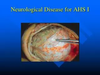
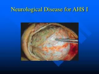
Neurological Stressors I-IV NUR240 Joy Borrero, RN, MSN
Assessment • Family history and genetic risk • Personal history • Level of consciousness and orientation • Memory: remote or long term, recall, immediate • Attention • Language and copying
Assessment • Cognition • Cranial nerves • Sensory function • Pain and temperature • Touch • Abnormal sensory findings
Assessment of Motor Function • Muscle strength • Cerebral or brainstem integrity • Abnormal motor findings
Assessment of Cerebellar Function • Coordination • Gait and equilibrium
Rapid Neurologic Assessment • Glasgow Coma Scale • Response to painful stimuli • Level of consciousness • Decortication • Decerebration • Pupil assessment
Laboratory Tests • Blood cultures necessary • Skull and spine x-ray tests • Cerebral angiography • CT scan: possible use of contrast medium, assess for allergic response, fluids • Positron emission tomography
Lumbar Puncture • Insertion of spinal needle into the subarachnoid space (between the third and fourth lumbar vertebrae) • Contraindicated in clients with increased intracranial pressure • Empty bladder • Position • Spinal headache possible from spinal tap
Electroencephalography (EEG) • Graphically records the electrical activity of the cerebral hemispheres • Sleep deprivation requirement • Anticonvulsants possibly withheld
Neurological Assessment of Intracranial Pressure • Normal ICP= <15mm Hg Increased intracranial pressure: • Causes and mechanisms: Increases in intracranial blood volume Increases in cerebral spinal fluid volume (CSF) Increases in the bulk of brain tissue-swelling
ICP • Signs & symptoms of increased ICP • Changes in LOC • Visual • Motor • Headache • Vomiting • Restlessness
ICP Cushing’s triad: • Increased MAP (D +1/3 S-D) • Increased pulse pressure (normally 40-50) • Bradycardia
Causes of Increased ICP • Increase in blood volume • Blood clot, edema, increased cerebral blood flow • Increased BP, PaCO2 • Decreased PaO2 • Vasodilitation- NTG, Nitroprusside • Increased intrathoracic pressure • Impairment of Cerebral Venous return
Compensatory Mechanism for Increased ICP • Displacement and reduction of volume of CSF- increased absorption by arachnoid villi • Reduction of volume of blood by vasoconstriction • Displacement of the tissues of the brain
Increased ICP • Treat pain, restrict fluid • Monitor patient • Cluster nursing care to promote rest Moderate increase in ICP (20-30): • Hyperventilate, maintain pCO2 25-30. • Sedate, neuro checks. Severe Increase in ICP over 40: • Constant hyperventilation, mannitol, barbituate coma (peds).
ICP Monitoring • Facilitates continual assessment of ICP and is more precise than relying on vague parameters • Devices include the intraventicular catheter, subarachnoid screw or bolt and epidural monitor. • ICP levels should be <15mm Hg • Assess client frequently for increasing ICP • Assess device insertion site for s/s infection
Management of Patients with Increased ICP Altered Cerebral Tissue Perfusion: • Decrease brain edema- osmotic diuretics, Lasix, Steroids • Control Temp • Lower CSF volume- venticulostomy drain • Positioning- avoid Trendelenberg and extreme hip flexion
Management of Patients with Increased ICP 5. Decrease blood volume Hyperventilation leads to resp alkalosis-vasoconstriction 6. Reduce cellular metabolic demands- Barbituates/ Barbituate Coma 7. Nursing implications of ICP monitoring
Neurosurgery-Craniotomy • Indications • Surgical Approaches: 1. Supratentorial 2. Infratentorial 3. Transphenoidal 4. Burr holes
Pre-op Craniotomy • Anticonvulsants • I & O, foley • Head shave prior to sx • Bowel prep only as ordered • Explain procedure and ICU course • Antiembolism stockings • No narcotics or hypnotics
Post-op Craniotomy • Adequate respiratory ventilation • Arterial pressure monitoring • Evaluate for cerebral edema and increasing ICP • Temp control • Medicate for HA and anticonvulsant meds: Dilantin (phenytoin) and Valium (diazepam) • Prevent aspiration • Prevent complications • Evaluate dsg for bleeding or leakage of CSF • Stool softeners
Intervention Don’ts: • Suction nose • Lower HOB • Restrain • Take rectal temp • Heavily sedate patient • Administer narcotics unless Double Checked
Post-op Transphenoidal Approach • PC Hemorrhage: Check nasal packing and frequent swallowing • Risk for infection- antimicrobials • Pain analgesics • Steroids • Assess visual acuity • Oral care q4h • HOB up, do not blow nose, bend or strain • Monitor for CSF leakage, postop meningitis, SIADH • Vaporizer and HOB up for 2 weeks postop
Head Injury- Skull fracture • Assessment • Fracture of the base of the skull nose- cerebrospinal fluid-rhinorrhea ears- cerebrospinal fluid-otorrhea • Ecchymosis or bruising seen over the mastoid ( Battle’s sign) • Bloody spinal fluid
Traumatic Brain Injury • Result of an external force applied to the head and brain causing disruption of physiologic stability • Diffuse axonal injury (shearing injuries)
Primary Brain Injury • Open head injury occurs when there is a skull fracture or when the skull is pierced by a penetrating object; the integrity of the brain and the dura is violated and exposure to outside contaminants occurs. • Closed head injury is the result of blunt trauma; the integrity of the skull is not violated.
Open Head Injury(Continued) • Comminuted fracture: involves fragmentation of the bone, with depression of bone into brain tissue • Open fracture: scalp is lacerated, creating a direct opening to brain tissue
Basilar Skull Fracture • Occurs at the base of the skull • Usually extends into the anterior, middle, or posterior fossa and results in cerebrospinal fluid leakage from the nose or ears • Potential for hemorrhage, damage to cranial nerves, and infection
Types of Closed Head InjuriesTBI- Traumatic Brain Injury • Mild concussion-transient disorder with momentary LOC followed by complete recovery • Contusion (coup and contrecoup injury)- bruising without tearing of vessels, results of brain hitting the skull • Laceration
Head trauma • Concussion- jostling brain • Contusion= bruising • Increased intracranial pressure- change in LOC first seen, lethargy, slowed speech, delayed responses, later change in VS • Establish airway, hyperoxygenate. • Proper positioning, HOB 30 degrees. • Minimal noise and stimulation
Types of Force • Acceleration injury is caused by an external force contacting the head, suddenly placing the head in motion. • Deceleration injury occurs when the moving head is suddenly stopped or hits a stationary object.
Brain Injury Complications • Increased ICP • Hemorrhage • Epidural hematoma • Subdural hematoma • Intracerebral hemorrhage • Loss of autoregulation • Hydrocephalus • Herniation
Epidural Hematoma • Occurs between the skull and the dura • Generally an arterial bleed • Classic picture- pt unconscious immediately after head trauma, awakens lucid and then lapses into coma • Goal is to relieve pressure and decrease ICP
Subdural Hematoma • Collection of blood between dura(outer meninges and arachnoid (middle layer meninges) • Generally venous • Acute- occurs within 24 hrs of injury • Subacute- symptomatic 2-14 days later • Chronic- weeks to months later • S&S depends on size of vessel involved and amount of bleeding • Management: Burr holes and craniotomy to remove clot
Subarachnoid Hemorrhage (SAH) • Bleeding into the subarachnoid space • Most common cause is rupture of aneurysm • Berry aneurysm- congenitally associated with smoking, ¼ die before reaching ER • More common in females • + Kernig’s sign • + Brudzinski reflex
SAH Clinical Manifestations • Change in LOC • Sudden severe HA • Photophobia • Nuchal rigidity • Low back pain • N & V • Fever, elevate dWBCs • Cranial nerve deficits • Motor weakness • EKG changes-bradycardia, AV block, PVCs
SAH TREATMENT • Surgery- metal clip for aneurysms • Amicar • Antifibrinolytics- controversial • Nimodipine (Nimtop)- to reduce risk of vasospasm • Phenytoin (Dilantin)- to tx seizures
Intracerebral Hemorrhage • Bleeding directly into brain tissue • CAUSES ARE SIMILAR TO SUB/EPIDURAL HEMATOMA • Tx- surgical- high mortality • Head trauma that compromises the function of the pituitary gland may lead to DI and SIADH
Head Trauma • Factors that predict a poor outcome: The presence of an intracranial hematoma Increasing age of the patient Impaired or absent eye movements or pupil light reflexes Abnormal motor responses Early sustained hypotension Hypoxemia or hypercapnia ICP greater that 20mm Hg
Clinical Care following a Head Injury • Airway • Establish baseline data • Prevent aspiration • Cardiovascular complications • Skull and scalp injuries • Infection prevention • Prevention of straining • Maintain normothermia
Clinical Care following a Head Injury • Fluid and electolyte, acid-base maintenance • Monitor restlessness, pain and disorientation • Pain management with mild analgesics • Assess for seizures • Positioning in bed to prevent stasis • Stress ulcers • Rest- prevent complications of inactivity • Space activities to prevent increase in ICP
Tumors of the Nervous System • All intracranial tumors can be fatal, no room for expansion • Classification: A. Intracranial tumors: Primary Metastatic Granulomas B. Spinal tumors C. Tumors of the peripheral nerves
Types of Intracranial Tumors • Gliomas • Neuromas • Meningiomas • Various blood vewssel tumors • Developmental tumors • Miscellaneous
General Symptoms of Intracranial Tumors • Caused by generalized disturbances of cerebral function • HA • N&V-no relation to meals • Papilledema • Seizures • Dizziness and vertigo • Changes in mental status
Management of Brain Tumors • Surgical excision • Brain mapping with steriotatic approach • Brachytherapy • Gamma knife for radiosurgery • Radiation • Chemotherapy • Mannitol to allow more chemotherapy across blood brain barrier • Thalidomide- decrease vascular supply • Growth Factor inhibitor tx- shrink tumor size • Decrease ICP • Assess for seizure activity and early signs of motor function impairment
Intracranial Aneurysms • Berry aneurysm is most common congenital aneurysm • Aneurysms result from: - a congenital defect in the middle layer of the vessel wall - degenerative changes in the vessel wall at the same site - constant stress caused by the force of the flow of blood particularly at the bifurcattion
Symptoms of Intracranial Aneurysm • Caused by compression of surrounding brain tissue or cranial nerves • Usually sudden onset of symptoms • Severe HA (occipital) and usually accompanied by vomiting • May lose consciousness immediately or initially present with confusion, lethargy • Generalized seizures • Meningeal irritation, stiff neck and back and leg pain, motor weakness, monoparesis, hemiparesis
Management • Surgical-evacuation of hemorrhage if life is threatened by increased pressure • Medical- antihypertensives antifibrinolytics decrease ICP prophylactic anticonvulsants