WRIST COMPLEX
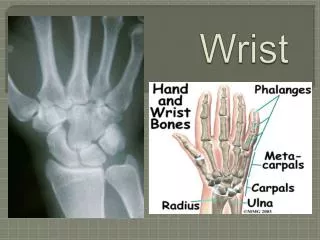
WRIST COMPLEX. Bones and Joints of the Wrist. Proximal Row of Carpal Bones. Review- testable Scaphoid: Most lateral. Forms floor of anatomical snuff box. Most commonly fractured wrist bone. Fractures may compromise radial artery in snuff box. Articulates with radius.

WRIST COMPLEX
E N D
Presentation Transcript
WRIST COMPLEX Bones and Joints of the Wrist
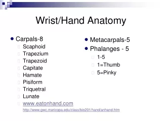
Proximal Row of Carpal Bones • Review- testable • Scaphoid: Most lateral. Forms floor of anatomical snuff box. Most commonly fractured wrist bone. Fractures may compromise radial artery in snuff box. Articulates with radius.
Proximal Row of Carpal Bones • Lunate: Articulates with radius • Triquetral: Articulates with ulna (via articular (ulnar) disc) during extreme ulnar deviation. • Pisiform: Sesamoid bone Forms in tendon of the flexor carpi ulnaris
Distal Row of Carpal Bones: • Trapezium: Most lateral • Trapezoid • Capitate • Hamate
Distal Row of Carpal Bones: • Entire complex enclosed in a common synovial membrane. • Articulations are plane joints that perform gliding motions.
Radiocarpal Joint • Condyloid (ellipsoidal) synovial joint. • Two degrees of freedom. • Articular surfaces: Scaphoid (convex) Lunate (convex) Distal radius: Two concave fossae (lateral and medial) Triquetral (convex) Only during extreme ulnar deviation
Radiocarpal Joint Ligaments • Lateral (radial) collateral ligament. • Medial (ulnar) collateral ligament. • Dorsal radiocarpal ligament. • Palmar radiocarpal ligament. • Strengthen capsule
Radiocarpal Joint Functions • Some flexion and extension • Ulnar deviation
Radiocarpal Joint Arteries • Articular arteries • Arise from dorsal and palmar carpal arches.
Radiocarpal Joint Nerves • Anterior interosseous branch of median nerve. • Posterior interosseous branch of radial nerve. • Dorsal and deep branches of the ulnar nerve.
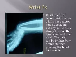
Radiocarpal Joint Injuries • Colle’s fracture • Scaphoid fracture Usually at “waist” Compromises radial artery in snuffbox
Midcarpal Joint • Made up of intercarpal joints: Between proximal and distal rows of carpals and between carpals. • Movements: Some flexion and extension. Radial deviation (abduction). Especially due to movement of head of capitate in its socket. • Enclosed within synovial capsule.
Midcarpal Joint • Ligaments: Dorsal ligaments. Palmar ligaments. Interosseous ligaments. • Nerves and arteries: Same as for radiocarpal.
Palmar Structure Sequence(radial to ulnar) • Radius • Radial artery • Flexor carpi radialis tendon • Median nerve: Under palmaris longus tendon
Palmar Structure Sequence(radial to ulnar) • Flexor digitorum superficialis tendons • Ulnar artery • Ulnar nerve • Flexor carpi ulnaris tendon
Carpometacarpal Joints • Plane synovial joints: • Motion: None for digits 2-3 Limited for 4 More mobile for 5
Carpometacarpal Joints • Saddle (sellaris) joint between metacarpus and trapezium: • Movements: Abduction/adduction Flexion/extension Circumduction Opposition
Metacarpophalangeal Joints • Condyloid synovial joints • Movements: Flexion/extension Abduction/adduction Some opposition at MCP 5 • Capsular ligaments: Palmar ligaments (pads) Collaterals
Interphalangeal Joints • Synovial hinge joints • Only flexion/extension allowed • Ligaments: Strong collaterals • Proximal interphalangeal joints (PIPs) • Distal interphalangeal joints (DIPs)
Dorsal Venous Drainage • Dorsal venous arch drains hand dorsum. • Medially drains into basilic. • Laterally drains into cephalic.
Lymphatic Drainage • Medial via lymph vessels accompanying basilic vein to: Supratrochlear nodes to: Lateral axillary nodes. • Lateral via lymph vessels accompanying cephalic vein to: Infraclavicular nodes to: Lateral axillary nodes.
Arterial Supply to Dorsum • Via dorsal arterial arch from: Radial and ulnar arteries. • Dorsal metacarpals. • Dorsal digitals.
Muscles of Dorsum of Hand • Long extensor tendons. • Dorsal interosseous muscles (4): Attachments: DAB: Abductors Middle finger is reference Middle finger has two First and fifth digits have none.
Superficial Palm • Palmar aponeurosis • Flexor retinaculum • Palmaris brevis
Palmar Aponeurosis • Triangular layer of deep fascia located between two eminences. • Provides protection for superficial vessels, nerves, and tendons. • Anchored to skin and flexor retinaculum. • Splits into four slips that blend with fibrous flexor sheaths of four medial digits (II – V).
Flexor Retinaculum • = Transverse carpal ligament. • Laterally attaches to tubercles of scaphoid and trapezium. • Medially attaches to hook of hamate and pisiform.
Palmaris Brevis Muscle • O: Flexor retinaculum and palmar aponeurosis. • I: Skin on medial side of palm. • A: Tenses skin on palm.
Carpal Tunnel Contents • Long flexor tendons of: Flexor digitorum superficialis Flexor digitorum profundus Flexor pollicis longus • Median nerve • Note: ulnar nerve and artery pass through Guyon’s canal.
Intrinsic Muscles of the Thumb • Thenar eminence: • Adductor pollicis: Innervation: Deep branch of ulnar nerve (C8, T1).
Thenar Eminence Muscles • Abductor pollicis brevis • Flexor pollicis brevis • Opponens pollicis • Innervation: Recurrent branch of median nerve (C8, T1).
Hypothenar Eminence • Intrinsic muscles for digit V. • Abductor digiti minimi • Flexor digiti minimi brevis • Opponens digiti minimi • Innervation: Ulnar nerve
Long Digital Flexors • Flexor digitorum superficialis • Flexor digitorum profundus
Flexor Digitorum Superficialis • Flexes PIP (and MCP and wrist). • Each tendon passes through fibrous flexor sheath. • Each tendon bifurcates opposite proximal phalanx. • Each tendon inserts on middle phalanx.
Flexor Digitorum Profundus • Flexes DIP (and PIP and MCP). • More active than superficialis. • Each tendon inserts on distal phalanx.
Vinculae • Small vascular bundles connecting palmar surface of phalanges with long flexor tendons. • Long and short
Dorsal Interossei • Four bipennate muscles. • Each arises via two heads from adjacent sides of two metacapals.
Dorsal Interossei • Insertion: Onto extensor expansions and: Radial sides of proximal phalanges 2 and 3; Ulnar sides of proximal phalanges 3 and 4. Note: digit has two dorsal interossei. • Abducts MP joints of digits 2-4: Reference is line through middle finger.
Palmar Interossei • Four unipennate muscles: First is sometimes considered part of flexor pollicis brevis. Supply each digit except third: Reference is middle finger. • Innervation for all interossei (incl. dorsal): Ulnar nerve
Lumbricals • Four small, narrow, elongated muscles. • Each arises from the radial side of a flexor digitorum profundus tendon. • Innervation: Two on radial side: Median nerve Two on ulnar side: Ulnar nerve • Flex MCP joints and extend IP joints.
Arterial Supply to Hand • Superficial palmar arch: Continuation of ulnar artery. • Deep palmar arch: Continuation of radial artery.
Route of Radial Artery • Smallest terminal branch of brachial artery. • Passes proximally deep to brachioradialis muscle. • Distally the artery lies against the radius lateral to the tendon of the flexor carpi radialis, where it can be felt (radial pulse). • Passes across scaphoid in anatomical snuff box.
Route of Radial Artery • Wraps around the dorsum of first metacarpus: Gives off arteries to the thumb and index finger. • Pierces the first dorsal interosseous muscle and reappears in the palm of the hand. • Gives rise to the deep palmar arch.