Download
1 / 40
410 likes | 571 Vues
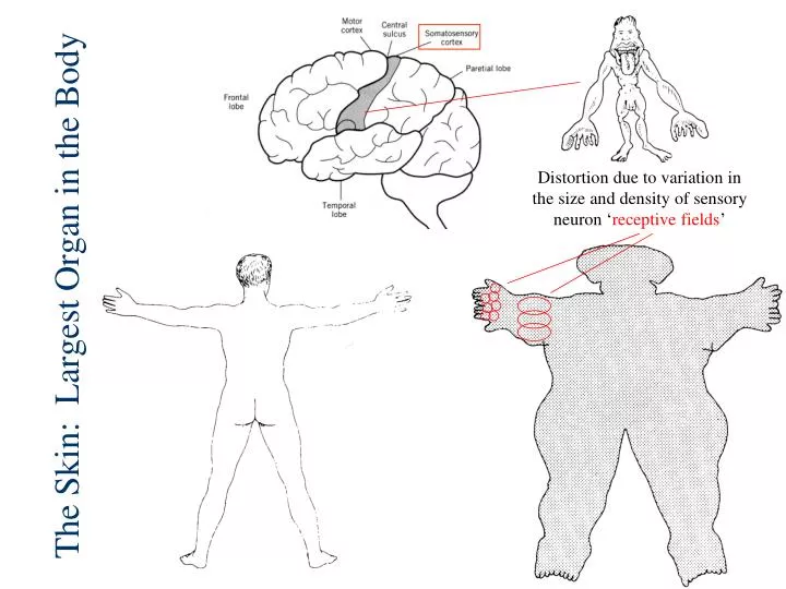
The Skin: Largest Organ in the Body. Distortion due to variation in the size and density of sensory neuron ‘ receptive fields ’. The Stimuli of Somatosensation. SKIN (body surface) Mechanical pressure: this is ‘touch’ Vibration (Hz): this is generally ‘texture’

E N D
The Skin: Largest Organ in the Body Distortion due to variation in the size and density of sensory neuron ‘receptive fields’
The Stimuli of Somatosensation • SKIN (body surface) • Mechanical pressure: this is ‘touch’ • Vibration (Hz): this is generally ‘texture’ • Damage/Temp (pain/hot/warm/cold) • Chemical (example is menthol) • MUSCLES &TENDONS (body position) • Stretch, Tension • Kinesthesis, Proprioception • BODY SURFACE + BODY POSITION = “HAPTIC PERCEPTION”
Free Nerve Endings • Receptors can be characterized in terms of: • Stimulation Type (pressure, vibration, temp, damage) • Size of Receptive Field (amount of branching) • Rate of Adaptation (slow, medium, fast) Basket Cell Pacinian corpuscle
Pacinian Corpuscles Detect Vibration (texture) Pressure Stimulus Pacinian Activity
Bone Red is motor Sensory fiber attached to tendon Receptors in Muscles and Tendons Encode Body Position Golgi Tendon Organ – Tension Receptor Blue is sensory Muscle Spindle – Stretch Receptor Muscle fiber Muscle
This is a Muscle spindle, but other Mechanoreceptors (i.e., Basket Cells and Pacinian Corpuscles) also work this way The cytoskeletal strands are like the ‘tip links’ of inner hair cells. Ion channels ‘pulled open’ by mechanical force.
Skin Senses: 2 Pathways to Cortex • Lemniscal Pathway (mechanorecepetors) • Tactile, pressure, Basket Cell (detect) • Tactile, vibration/texture, Pacinian Corpuscle (ID) • Spinothalamic Pathway (free nerve endings) • Tissue Damage, pain, Nociceptor (detect) • Temperature, hot/cold, Thermal Receptor (ID) • This pathway is ‘gated’ in the spinal cord
D o r s a l – S e n s o r y Thalamus V e n t r a l – M o t o r Hypothalamus Cortex frontal Cortex parietal • Thinking in 3D • Dorsal/Ventral Organization • Left/Right Crossing Thalamus D O R S A L Hindbrain Dorsal Ventral MUSCLES MUSCLES SENSORY NEURONS Ventral Dorsal Spinal Cord
Primary Cortex Thalamus 2D Receptor Array However…
Cortex Is ‘Modular’ Means the size and density of cortical columns is fixed Thalamus Dense 2D Receptor Array
Fuzzy Cortex frontal Cortex parietal Lemniscal System (mechanoreceptors) Thalamus Hindbrain Spinal Cord PACINIAN CORPUSCLE
Receptive fieldsperiphery vs. cortex Stimulation anywhere within this large receptive field goes to one cortical column Stimulation within this tiny receptive field goes to one cortical column
Two-Point Discrimination Less convergence= More discrimination, Higher threshold Converging neurons = Less discrimination, Lower threshold Detect Identify
The brain has no ‘sense’ of itself Sensory (parietal) Motor (frontal)
Where best for Braille? Two-Point Thresholds
Thresholds for Detection and Identification Pressure (Detect) Two-Point (ID)
Monkey Cortex
Experience Changes Cortical Maps • what happens if you lose a finger? • cortical maps will readjust • experience alone can readjust - the example of violin training - young vs. old
What is the result of all this ‘experience’? Smart? Dumb? Dumber?
Variation in Cortical Maps Overall brain weight differs by ~30% Size of primary cortical areas differs by as much as 100%
Normal Cortex is allocated based on use The beauty of modular architecture “Columns is Columns” Blind Touch Vision Audition
Pain Is A Perception: The Stimulus is Tissue Damage Pain Pain Pain Pain Nociceptors respond to AND release chemical stimuli (the basis of inflammation)
Heart Attack? Lung Pain? Arm Pain? Convergent Excitation: lower thresholds (better detection) come at a cost of lousy ID.
L-fibers are mechanoreceptors Spinal Gate Theory: S-fibers are free nerve endings 1. Two Ways to Inhibit 2.
Spinothalamic System (free nerve endings) Cortex frontal Cortex parietal OUCH! Thalamus 3. Hindbrain Spinal Cord 2. 1. Nociceptor(excitatory) 2. Mechanoreceptors (excitatory) 3. Modulatory Brainstem neurons (excitatory) Opiate Neuron (inhibitory) 1.
Spinothalamic System (free nerve endings) Cortex frontal Cortex parietal Thalamus 3. Hindbrain Spinal Cord 2. 1. Nociceptor(excitatory) 2. Mechanoreceptors (excitatory) 3. Modulatory Brainstem neurons (excitatory) Opiate Neuron (inhibitory) 1.
Spinothalamic System (free nerve endings) Cortex frontal Cortex parietal Thalamus 3. Hindbrain Spinal Cord 2. 1. Nociceptor(excitatory) 2. Mechanoreceptors (excitatory) 3. Modulatory Brainstem neurons (excitatory) Opiate Neuron (inhibitory) 1.
Midbrain (DA) Hindbrain (5-HT, NE) Architecture of ‘Modulatory’ Systems “one-to-many” Dopamine (DA) Serotonin (5-HT) Norepinephrine (NE)
Endogenous Opiates • Common practice to name neurotransmitters after the plant-derived chemical that mimics their action in the brain • Morphine-like neurotransmitters • Endorphins, Enkephalins • Role in suppressing pain (i.e., inhibiting input from nociceptors) • Opiate neurons found throughout brain and spinal cord – not all are involved in pain
EPSP: glutamate IPSP: gaba or endogenous opiate + YES! NO! - Synaptic potentials are brief (a few milliseconds) positive or negative changes in voltage. Occur at dendrites – positive and negative summate. Caused by neurotransmitter released from a presynaptic neuron. EPSPs increase likelihood of action potentials, IPSPs decrease that likelihood. Drug effect: excitatory agonist YEEEEEEEEEEEEEEEEEEEEEEEEEEEEEESSSSSSSSSSSSSSSSSSSSSSSSSS! Agonist drugs can also produce positive or negative changes in voltage. They do so by mimicking the chemical structure of naturally-occurring neurotransmitters. However, the DURATION of their effects can be minutes or hours. Neurons respond to this stimulation by making themselves more or less excitable. Leads to dose tolerance and with persistent use, chemical dependence. NOOOOOOOOOOOOOOOOOOOOOOOOOOOOOOOOOOOOOOO! Drug effect: inhibitory agonist
Why drugs produce chemical dependence. . . . . . even ‘good’ ones Short term effect of morphine is increase in K+ current Long term effect of morphine is increase in Na+ current
The ‘Spinal Gate’ in action Cell bodies in brainstem dorsal ventral
Activation of a Nociceptor: Inhibition by Enkephalin / Morphine
Cone Snail Venom Venom consists of a ‘cocktail’ of proteins One of these venom proteins blocks ‘N’ type calcium channels Nociceptors have ‘N’ type calcium channels on their axon terminals
Receptor Agonists / Antagonists Reuptake Inhibitors Neurotransmitters K+ Na+ Ca++ Where a venom (or drug) could work. . .