Mitogen-Activated Protein Kinase 1
240 likes | 511 Vues
Mitogen-Activated Protein Kinase 1. Emerlyn Goh. Overview. Introduction Structure Function Related Molecules Specific Aspect * References. Introduction. What is a kinase? Phosphorylation What are mitogen-activated protein kinases?

Mitogen-Activated Protein Kinase 1
E N D
Presentation Transcript
Mitogen-Activated Protein Kinase 1 Emerlyn Goh
Overview • Introduction • Structure • Function • Related Molecules • Specific Aspect* • References
Introduction • What is a kinase? • Phosphorylation • What are mitogen-activated protein kinases? • a.k.a MAPKs (microtubule-associated protein kinase/mitogen activated protein kinase), ERKs (extracellular signal-regulated kinases) • History • Signal transduction pathways • Highly Conserved • Transmembrane communication • MAP2K, MAP3K
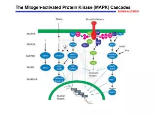
MAP Kinase Cascades Source: CALBIOCHEM et al, 2008
ERK1/2 Signal Transduction Pathway • ERK1/2 similar (ERK2 focus) • ERK2 = MAPK1 • Mitogen(EGF)Receptor (RTK)GTPase(ras)Protein kinase (raf)MAPK cascadecellular response • Often called “Ras-Raf-MEK-ERK” pathway • Mechanisms of activation of MAP3K largely unknown • Thr-575 important
Structure of ERK2 • Inactive form: Tyr-185 blocks active site • 2 domains further apart • Active form: Tyr-195, Thr-183 • Dual phosphorylation • Alignment of p-tyrosine & p-threonine, turn towards surface arginine-rich binding sites to maximize ATP binding • Local and global conformation changes • Active site closure
Unphosphorylated ERK2 Phosphorylated ERK2 PDB ID 1ERK (Zhang et al, 1997) and 2ERK (Canagarajah et al, 1998)
Structure Comparison: Activated/Inactivated Residues PDB ID 1ERK (Zhang et al, 1997) and 2ERK (Canagarajah et al, 1998)
Functions of ERK2 • Regulates proliferation and differentiation of post-mitotic cells • Development of the mesoderm/placenta in many eukaryotes* • Cell growth (ex. transcription factor Elk-1) • Phosphorylation of P53 in cervical/breast carcinoma cells by ERK2 suggests apoptosis • Cell cycle progression and cancer expression (ex. transcription factor c-myc)
Functions, Explained • Elk-1 : transcription factor involved in the expression of c-fos (Gille et al, 1995) • C-fos accounts for cell differentiation, growth • P53 : Breast carcinoma cells + doxorubicin activation of ERK2 phosphorylates P53 on Thr-55 residue suggests apoptosis in some cells and drug resistance in cancerous mutants (Pei et al, 2004) • C-Myc : proto-oncogene regulated by MAPK pathway • Overexpression pancreatic/colon cancers • Cyclin expression and G1-S progression • Cancer treatment? (Marampon et al, 2006)
Related Molecule: MKP3 • Dual specificity MAPK phosphatases (MKPs/DSPs) • MKP3 (DUSP6) prototypical ERK2-specific • Able to form stable complex with ERK2 • KIM peptide binds to N-terminal noncatalytic site opposite activation loop of ERK2 • Tightly bound for catalytic activation, dephosphorylation of ERK2 • Underexpression ERK2 overexpressionpancreatic carcinogenesis
MKP3 KIM PeptideComplexed with ERK2PDB ID 1fys (Liu et al, 2006)
Activation of MKP3 • Activation site is a shallow cleft to accommodate Tyr/Thr residues on ERK2 • Inactivated MKP3: Asp-262 found in loop 5.5Å away from active site • Once ERK2 binds: Asp-262 loop folds to bring aspartate into active site catalytically active stable conformation (Theodosiou et al, 2002)
Unactivated MKP3(PDB ID 1MKP Stewart et al, 1998) PDB ID 1MKP (Stewart et al, 1999)
How is MKP3 Regulated? • Posttranslational mechanism by ERK2 • Via MEK-ERK pathway MKP3 phosphorylated on Ser-159, Ser-197 • Phosphorylation on either serine enhances proteasomal degradation of MKP3 • Experiment: Double mutations on the serines caused 3-fold increase in MKP3 half-life • This is a positive feedback loop by ERK2 by promoting degradation of one of its main inhibitors (Marchetti et al, 2005)
Related Molecule: Aurora-A Kinase • AURKA gene • Key roles in cell mitosis • Potential oncogene, uncertain • Pancreatic cancer linked to hyperactivation and overexpression of Aurora-A by MAPK pathway • Drug treatment options? • (Fu et al, 2007)
Aurora-A Phosphorylated/Unphosphorylated Compared • Unactive Thr exposed for phosphorylation • Phosphorylated Thr pulls inward to a buried positionactive conformation PDB ID 1OL7 Bayliss et al, 2003 and PDB ID 1MQ4 Nowakowski et al, 2003
Essential Role of ERK2 in Mesodermal and Placental Development • Experiment: ERK2-deficient mice embryos placental development problems • ERK2-deficient mice poor fetal vascularization in placentasevere defect in labyrinthine layer of placentapoor development • As a result fetuses exhibited extreme growth retardation, thin heart walls, and death (Hatano et al, 2003) • Experiment: ERK2 mutant embryos failed to develop mesoderm, defective differentiation (Li et al, 2003)
Codon 99 Mutant of ERK2 Wildtype, Heterozygous, and Homozygous compared-Arrowhead=fetal nucleated erythrocytes-Arrows=mother nucleated erythrocytes-Heterozygoushypomorphic alleleHatano et. al, 2003
References • Gille et al, 1995: http://www.ncbi.nlm.nih.gov/pubmed/8548291 • Pei et al, 2004: http://www.nature.com/onc/journal/v23/n20/full/1207426a.html#bib41 • Waas et al, 2007: http://www.ncbi.nlm.nih.gov/pubmed/17251036?ordinalpos=1&itool=EntrezSystem2.PEntrez.Pubmed.Pubmed_ResultsPanel.Pubmed_RVDocSum • Theodosiou et al, 2002: http://www.pubmedcentral.nih.gov/articlerender.fcgi?artid=139386 • Marchetti et al, 2005: http://www.ncbi.nlm.nih.gov/pubmed/15632084 • Fu et al, 2007: http://mcr.aacrjournals.org/cgi/content/full/5/1/1?ck=nck • Hatano et al, 2003: http://www.genestocellsonline.org/cgi/reprint/8/11/847.pdf • Stewart et al, 1998: PDB ID 1MKP Structure of MKP3 • Liu et al, 2006: PDB ID 1FYS • Ohren et al, 2004: PDB ID 1s9j • Krysan, http://www.hort.wisc.edu/Krysan/) • Zhang et al, 1997: PDB ID 1ERK • Canagarajah et al, 1998: PDB ID 2ERK • Calbiochem et al, 2008: http://www.emdbiosciences.com/html/CBC/phosphorylation_inhibitors_mitogen-activated_protein_kinase.htm • Li et al, 2003: http://www.pnas.org/cgi/content/abstract/100/22/12759 • Marampon et al, 2006: http://www.molecular-cancer.com/content/5/1/31