P = Flow x resistance
150 likes | 392 Vues
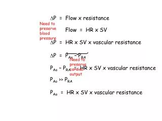
P = Flow x resistance. Need to preserve blood pressure. Flow = HR x SV. P = HR x SV x vascular resistance. P = P Ao – P RA. Need to preserve cardiac output. P Ao – P RA = HR x SV x vascular resistance. P Ao ›› P RA. P Ao = HR x SV x vascular resistance. Decreasing

P = Flow x resistance
E N D
Presentation Transcript
P = Flow x resistance Need to preserve blood pressure Flow = HR x SV P = HR x SV x vascular resistance P = PAo – PRA Need to preserve cardiac output PAo – PRA = HR x SV x vascular resistance PAo ›› PRA PAo = HR x SV x vascular resistance
Decreasing compliance Compliance Volume Pressure
Decreasing contractility Contractility Pressure Volume
Contractility and compliance Systole and diastole Systolic Decreasing contractility Decreasing compliance Pressure Diastolic Volume
Normal pressure volume loop The ventricle continues to empty until the end of the action potential. When the ventricular pressure exceeds the aortic diastolic pressure, the aortic valve opens and the ventricle starts emptying into the aorta. PAo systolic PAo diastolic The ventricle is depolarised and contracts (closing the mitral valve). Pressure LVEDP The ventricle fills passively during diastole. Upon repolarisation, the ventricle relaxes, resulting in closure of the aortic valve and opening of the mitral valve. SV Volume
Decreased preload volume depletion PAo systolic LVEDP SV
Decreased compliance pericardial constriction/tamponade restrictive/hypertrophic cardiomyopathy myocardial ischemia PAo systolic Pressure LVEDP SV Volume
Contractile dysfunction Acute myocardial infarction/ischemia PAo systolic LVEDP SV
SV Contractile dysfunction Dilated cardiomyopathy PAo systolic LVEDP SV
Increased afterload Hypertension PAo systolic LVEDP SV
Decreased preload and decreased afterload sepsis excessive vasodilatation PAo systolic Pressure LVEDP Volume
contractility afterload compliance Impaired preload shock pulmonary edema Normal Stroke volume LVEDP