Download
1 / 35
490 likes | 1.2k Vues
Antiarrhythmic Drugs. Drug List. Physiology Review. To function efficiently, heart needs to contract sequentially (atria, then ventricles) and in synchrony Relaxation must occur between contractions

E N D
Physiology Review • To function efficiently, heart needs to contract sequentially (atria, then ventricles) and in synchrony • Relaxation must occur between contractions • Coordination of heartbeat is a result of pacemaker activity of the SA node and transfer of impulses through AV node and the purkinje system
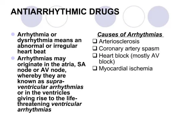
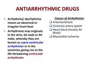
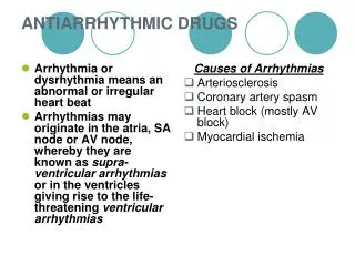
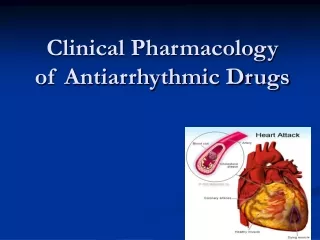
Arrhythmia A condition where there is a disturbance in • Pacemaker impulse formation • Impulse conduction • Combination of the two Results in change of heart rate or contraction of heart muscle in a way that is insufficient to maintain normal cardiac output To understand how antiarrhythmic drugs work, need to understand electrophysiology of normal contraction of heart
Ventricular Arrhythmia • Ventricular arrhythmias are common in most people and are usually not a problem but… • VA’s are most common cause of sudden death • Majority of sudden death occurs in people with neither a previously known heart disease nor history of VA’s • Medications which decrease incidence of VA’s do not decrease (and may increase) the risk of sudden death treatment may be worse then the disease!
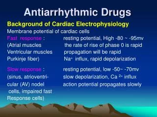
Electrophysiology - resting potential • A transmembrane electrical gradient (potential) is maintained, with the interior of the cell negative with respect to outside the cell • Caused by unequal distribution of ions inside vs. outside cell • Na+ higher outside than inside cell • Ca+ much higher ““““ • K+ higher inside cell than outside • Maintenance by ion selective channels, active pumps and exchangers
Contraction of ventricles ECG (EKG) showing wave segments Repolarization of ventricles Contraction of atria
Cardiac Action Potential • Divided into five phases (0,1,2,3,4) • Phase 4 - resting phase (resting membrane potential) • Phase cardiac cells remain in until stimulated • Associated with diastole portion of heart cycle • Addition of current into cardiac muscle (stimulation) causes • Phase 0 – opening of fast Na channels and rapid depolarization • Drives Na+ into cell (inward current), changing membrane potential • Transient outward current due to movement of Cl- and K+ • Phase 1 – initial rapid repolarization • Closure of the fast Na+ channels • Phase 0 and 1 together correspond to the R and S waves of the ECG
Cardiac Action Potential • Phase 2 - plateau phase • sustained by the balance between the inward movement of Ca+ and outward movement of K + • Has a long duration compared to other nerve and muscle tissue • Normally blocks any premature stimulator signals (other muscle tissue can accept additional stimulation and increase contractility in a summation effect) • Corresponds to ST segment of the ECG. • Phase 3 – repolarization • K+ channels remain open, • Allows K+ to build up outside the cell, causing the cell to repolarize • K + channels finally close when membrane potential reaches certain level • Corresponds to T wave on the ECG
Differences between nonpacemaker and pacemaker cell action potentials • Pacemaker Cells - Slow, continuous depolarization during rest • Continuously moves potential towards threshold for a new action potential (called a phase 4 depolarization)
Mechanisms of Cardiac Arrhythmias • Result from disorders of impulse formation, conduction, or both • Causes of arrhythmias • Cardiac ischemia • Excessive discharge or sensitivity to autonomic transmitters • Exposure to toxic substances • Unknown etiology
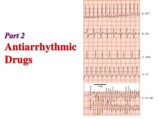
Types of arrhythmias Tachyarrythmias Bradyarrhythmia Tachyarrythmias Atrial: A fibrillation, Aectopics, Atrial tachycardia Nodal: Supraventricular Tachycardia Sinus Tachycardia ventricular: Ventricular Tachycardia , ventricular ectopics,Ventricular fibrillation or flutter
Brady arrhythmia: • Heart rate is less than 60 except in ( athletes) • Heart block • Sinus brady -cardia
Antiarrhythmic drugs • Biggest problem – antiarrhythmics can cause arrhythmia! • Example: Treatment of a non-life threatening tachycardia may cause fatal ventricular arrhythmia • Must be vigilant in determining dosing, blood levels, and in follow-up when prescribing antiarrhythmics
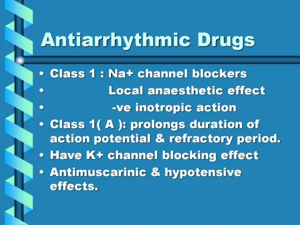
Classification of Antiarrhythmic Drugs Class I: Sodium channel blockers (membrane-stabilizing agents) 1 a:Block Na+channel and prolong action potential 1 b: Block Na+channel and shorten action potential1 c:Block Na + channel with no effect on action potential Class II: β- blockers Class III: Potassium channel blockers (main effect is to prolong the action potential) Class IV: Slow (L-type) calcium channel blockers
Class 1a Drugs • They are the oldest group of antiarrhythmic drugs and are still widely used. Procainamide • Blocks sodium channels, slows the upstroke of the action potential, slows conduction, prolongs the QRS duration of the ECG. • The drug also prolongs the action potential duration by nonspecific blockade of potassium channels. • Procainamide has direct depressant actions on sinoatrial and atrioventricularnodes
Procainamide Extracardiac Effects Procainamide has ganglion-blocking properties. This action reduces peripheral vascular resistance and can cause hypotension, particularly with intravenous use. Pharmacokinetics: Absorbed well orally. Metabolized in liver ,portion of the drug can be acetylated to NAPA which has little class 3 activity it is excreted by kidney . Toxicity : QT interval prolongation, and induction of torsade de pointes arrhythmia and syncope. New arrhythmias can be precipitated. Lupus erythmatosus, Pleuritis , pericarditis, or parenchymal pulmonary disease. Nausea and diarrhea (in about 10% of cases), rash, fever, hepatitis (< 5%) Therapeutic Use : Therapeutic Use Atrial and ventricular arrhythmias
Quinidine Similar to procainamide. Also blocks K+ channels. Adverse effects Anticholinergic(i.e. anti-muscarinic), blocks α receptors. Diarrhea , nausea A syndrome of headache, dizziness, and tinnitus (cinchonism) is observed at toxic drug concentrations. Excessive QT interval prolongation and induction of torsade de pointes arrhythmia. Toxic concentrations of quinidine also produce excessive sodium channel blockade with slowed conduction throughout the heart. Not very commonly used these days and largely replaced by calcium antagonists in clinical practice
Class 1b Drugs Lidocaine Block Na + channel and shorten action potential duration. Lidocaine has a low incidence of toxicity. A high degree of effectiveness in arrhythmias associated with acute myocardial infarction. It is used only by the intravenous route. Cardiac Effects Lidocaine blocks activated and inactivated sodium channels with rapid kinetics it shorten the period of repolarization (phase 3) thereby the period of action potential ,it has no effect in refractory period . Adverse effects: Lidocaine is one of the least cardiotoxic of the currently used sodium channel blockers .it shows little impaiment to left ventricular function and no negative iontropic effect,Paresthesias, nausea of central origin, lightheadedness, hearing disturbances. In preexisting heart failure, lidocaine may cause hypotension
Class 1b Drugs Pharmacokinetics: Extensive first-pass hepatic metabolism Lidocaine must be given parenterally ,Lidocaine has a half-life of 1–2 hours. Therapeutic Use: Lidocaine is the agent of choice for termination of ventricular tachycardia and prevention of ventricular fibrillation after cardioversion in the setting of acute ischemia (Myocardial infarction)
Mexiletine (1b) Mexiletine is given orally. Its electrophysiologic and antiarrhythmic actions are similar to those of lidocaine. Therapeutic use: Treatment of ventricular arrhythmias associated with pervious MI. Pain due to diabetic neuropathy and nerve injury. Adverse effects: Tremor , blurred vision, and lethargy. Nausea
Tocainde (1b) • Similar to Mexilitine and Lidocaine • It is used for treatment of ventricular tachy- arrhythmias • Can lead to plumonary fibrosis
Phenytoin (1b) • It is an other agent in this category and is usually used to treat ventricular arrhythmias caused by cardiac glycosides
Class 1c Drugs Flecainide Blocks Na+ channel with no effect on action potential. Flecainide is a potent blocker of sodium and potassium channels. (Note that although it does block certain potassium channels, it does not prolong the action potential or the QT interval). It has no antimuscarinic effects, it prolong the effective refractory period. It suppress the upstroke of phase 0 in purkinji fibers and all myocardial tissues It shows depression of conduction in all cardiac cells Automaticity is reduced by an increase in threshold potential It has prominent effect even in normal heart Recent data have cast serious doubt about safety of this class
Class 1c Drugs Pharmacokinetics:Flecainide is well absorbed and has a half-life of approximately 20 hours. Elimination is both by hepatic metabolism and by the kidney Therapeutic Uses: Premature ventricular contractions. Atrial fibrillation (AF), Wolf Parkinson White (WPW) syndrome Toxicity: Arrhythmia, can aggravate congestive heart failure because it has a negative ionotropic effect
Class 1c Drugs • Propafenone This drug is similar to Flecainide, slows conduction in all cardiac cells and is considered broad spectrum anti-arrhythmic agent.
Class 2 Drugs. Beta Blockers PROPRANOLOL:Suppress adrenergically mediated ectopic activity. Drugs have antiarrhythmic properties by virtue of their β -receptor–blocking action and direct membrane effects Some of these drugs have selectivity for cardiac β 1 receptors e.g. metoprolol, Some have intrinsic sympathomimetic activity e.g. pindolol.Some have marked direct membrane effects, and some prolong the cardiac action potential These agents can prevent recurrent infarction and sudden death in patients recovering from acute myocardial infarction.
Class 2 Drugs. Beta Blockers Therapeutic Uses:Sinus tachycardia, Atrial/ nodal extrasystole. Pheochromacytoma. Arryhthmia due to halothane /digitalis. WPW syndrome Esmololis a short-acting β blocker used primarily as an antiarrhythmic drug for intraoperative and other acute arrhythmias Sotalolis a nonselective β -blocking drug that prolongs the action potential.
Class 3 Antiarrhythmic DrugsAmiodarone Amiodaronemarkedly prolongs the action potential duration (and the QT interval on the ECG) Amiodarone also significantly blocks inactivated sodium channels. Amiodarone also has weak anti-adrenergic and calcium channel blocking actions ExtracardiacEffectsAmiodarone causes peripheral vasodilation.
Pharmacokinetics Incompletely and slowly absorbed orally. It undergoes hepatic metabolism, and the major metabolite, desethylamiodarone, is bioactive. The elimination half-life is complex. The drug accumulates in many tissues, including the heart (10–50 times more so than in plasma), lung, liver, and skin, and is concentrated in tears Therapeutic Use: Recurrent ventricular tachycardia, atrial fibrillation. Rapid termination of supraventricular tachycardia (SVT), ventricular tachycardia (VT) and WPW syndrome Works on both supraventricular and ventricular arrhythmias.
Amiodarone adverse effects • Fatal pulmonary fibrosis • Abnormal liver function tests and hepatitis • Photodermatitisand a gray-blue skin discoloration in sun-exposed areas (smurf skin) • Corneal micro-deposits and discoloration • Optic neuritis: Halos develop in the peripheral visual fields, may progress to blindness • Hypothyroidism or hyperthyroidism • Bradycardia and heart block
Class 4 Antiarrhythmic DrugsCalcium Channel-Blocking Drugs Verapamil: Cardiac Effects.Verapamil blocks both activated and inactivated L-type calcium channels. AV nodal conduction time and effective refractory period are prolonged. Slows the SA node by its direct action Extracardiac Effects.Peripheral vasodilation (Less than nifedipine) Pharmacokinetics:Absorbed orally. It is extensively metabolized by the liver Therapeutic Use.Supraventricular tachycardia. Atrial fibrillation and flutter
Miscellaneous Antiarrhythmic Agents These include digitalis, adenosine, and magnesium Adenosine Mechanism of Action: Adenosine is a nucleoside that occurs naturally throughout the body. Its half-life in the blood is less than 10 seconds. Activation of acetylcholine sensitive K + channels and inhibition of calcium current. Atrioventricular nodal conduction and the atrioventricular nodal refractory period. It is drug of choice (DOC) for conversion of paroxysmal supraventricular tachycardia to sinus rhythm. High efficacy (90–95%) and very short duration of action. Adverse effects: Flushing, shortness of breath or chest burning, high-grade atrioventricularblock. Atrial fibrillation may occur. Headache, hypotension, nausea, and paresthesias
Magnesium Mechanism of action is not known. USES: Digitalis -induced arrhythmias. Torsade de pointes even if serum magnesium is normal.
Pacemakers • Surgical implantation of electrical leads attached to a pulse generator • Over 175,000 implanted per year • Leads are inserted via subclavian vein and advanced to the right side of the heart • Two leads used, one for right atrium, other for right ventricle • Pulse generator containing microcircuitry and battery are attached to leads and placed into a “pocket” under the skin near the clavicle • Pulse generator sends signal down leads in programmed sequence to contract atria, then ventricles • Pulse generator can sense electrical activity generated by the heart and only deliver electrical impulses when needed. • Pacemakers can only speed up a heart experiencing bradycardia, they cannot alter a condition of tachycardia