Download
1 / 21
220 likes | 1.53k Vues
Digestive system III PEDIATRIC LIVER DISEASES . Professor shahenaz M. Hussien. Manifestations of liver diseases. Symptoms: Jaundice with dark colored urine and pale stool. Abdominal distension; due to hepatomegaly. Bleeding tendency e.g. hematemesis, melena and epistaxis.

E N D
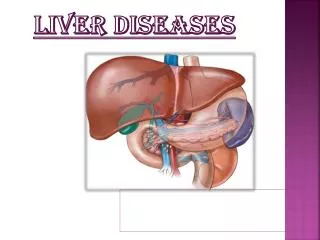
Digestive system III PEDIATRIC LIVER DISEASES Professor shahenaz M. Hussien
Manifestations of liver diseases Symptoms: • Jaundice with dark colored urine and pale stool. • Abdominal distension; due to hepatomegaly. • Bleeding tendency e.g. hematemesis, melena and epistaxis. • Purities. • Failure to thrive and malaise. • Abdominal pain, anorexia, fever and vomiting in cases of hepatitis. • Metabolic consequence of liver diseases e.g. encephalopathy. Signs: • Enlarged or shrunken liver. • Signs of portal hypertension; splenomegaly, dilated abdominal wall veins. -Ascites. -Clubbing especially in chronic liver disease.
Investigations of liver diseases Liver function tests I. evidence of cellular injury: assessed by Serum transaminases: • Alanineaminotransferase (ALT) normal value is 5-45U/L • Aspartatetransaminase (AST) normal value 15-55U/L • They are increased in liver necrosis marked in acute hepatitis. ALT is a more sensitive indicator of liver damage. II. Excretory function of the liver: can be assessed by:- • Bilirubin level: normally no bilirubin in urine. It is present in Obstructive and Hepatocellular jaundice • Urobilinogen in urine is increased in: -Hepatocelluar jaundice. -Hemolytic anemia. • Serum 5 nucleotidase is raised in intrahepaticcholestasis. • Increased S. gamma glutamyltransferase in obstructive jaundice.
Decreased serum albumin level in liver cirrhosis. • Elevated Serum immunoglobulins in chronic liver diseases. • Delayed prothrombin time and concentration (normal value 10-13 seconds and 85%-100% respectively). Imaging study of the liver:- • Liver ultrasound it shows: • Diffuse liver disease; cirrhosis, metabolic disease, hepatic periportalbilharzial fibrosis and fatty liver. • Biliary channel patency, choledocal cyst and gall stones. • Focal lesions, liver abscess - Ascites and subphrenic abscess. • Radioisotopic scanning it display liver size, morphology, focal lesions and biliary excretion. • Computerized tomography (CT) it demonstrates the presence of focal lesion, and cyst. • Magnetic resonance imaging (MRI) it can shows patency of biliary tree and focal lesion. • Liver biopsy and histopathological examination, it is the key for diagnosis of most liver disease.
HEPATOMEGALY Definition:- It is an inflammatory process of the hapatocytes characterized by degeneration and regeneration with loss of hepatic architecture. Types:- • Acute: less than six months duration • Chronic: more than six months Etiology: I- Infections, which may be:- Viral: • Hepatotropic viruses e.g A, B, C, D, E, F, G, H viruses • Non -hepatotropic: infect the liver in the course of other systemic illness: - Epstein-Barr virus (EBV). -Cytomegalovirus (CMV). - Coxsakie, -ECHO, -rubella, -varicella and measles viruses.
Bacterial : - As a part of generalized septicemia. - Isolated pyaemic liver abscess. - Leptospirosis. Protozoal: e.gamoebic hepatitis. II- Drugs and Toxins: • Anti T.B. e.gIsoniozid - antimetabolites - anticonvulsant:- valporic acid • Irradiation - chloropromazin - total parenteral nutrition Carbontetrachloride - halothane III- Immunological Disorders -As apart of : S.L.E and J.R.A - Isolated auto immune hepatitis IV- Metabolic Causes: • Alpha antitrypsine deficiency - galactosemia - tyrosenemia • Haemosidrosis - Wilson disease V- Vascular Causes - Hepatic vein thrombosis. - Hepatic artery thrombosis VI- Tumors: - Primary: hepatoma or hepatoblastoma - Secondary:- neuroblastoma, lymphoma, leukemia
HEPATITIS -A Mode of transmission: fecal-oral route Incubation period: 15-45 days (average 4 week). The virus is excreted in stool during the first few weeks of infection, prior to the onset of symptoms. Clinical manifestations: Acute Viral Hepatitis: 1- Prodromal stage: (1-2 weeks)(pre-Icteric) • Headache, anorexia, malaise. • Nausea, vomiting, lethargy. This phase can be passed un-noticed by the parents. 2- Icteric phase (2-3 weeks): Characterized by: • jaundice - dark urine. • Abdominal pain – soft enlarged, and tender liver. 3- Convalescence phase (1-2 weeks) - After which the child become nearly normal. In endemic areas 30-80 % of children acquire subclinical or anicteric infection in the first few years of life. -Anorexia - Nausea – Vomiting -Fever - Abdominal discomfort -Irregular bowel motions for a few days - Dark urine and mild scleral jaundice.
Laboratory diagnosis • Liver function tests shows:- • Raised serum level of .direct and indirect bilirubin Urine contains both bilirubin ( dark color) and Urobilinogen. • Marked elevation of serum transaminases (ALT and AST). • Increase in serum levels of alkaline phosphatase and 5 nucleotidase. IgM Anti- HAV is detected at the onset of the symptoms and disappears within 4 weeks while IgG anti-HAV persists for life Complication Acute fulminant hepatitis. • It is a rare condition with massive destruction of the liver cells. • It is presented clinically by persistent vomiting, disorientation, encephalopathy, bleeding tendency, edema and ascites. Aplastic anemia • Is a very rare complication it is transient but may be fatal. • It is due to bone marrow depression. • Death is usually due to serious infection due to depressed immunity Cholestassis • The patient becomes intensely pruritic and jaundiced • It is due to hepatocyte edema which may cause element of obstruction
Treatment • There is no specific therapy for acute viral hepatitis, • Most children are managed at home except if liver cell failure is suspected. • Balanced diet with low fat intake should be given. Prevention • Period of infectivity: Contagious for about 7 days before and 7 days after the onset of jaundice. • Period of isolation from school: From the appearance of dark color urine and appearance of jaundice till 7 days after disappearance of jaundice. • Careful hand washing after changing diapers and before preparation of food. -Fly control. • Intramuscular immunoglobulin may be indicated in pre and post exposure. • Hepatitis A vaccine is now available to be given to children. Contacts are immunized with immunoglobulin or the vaccine.
HEPATITIS -B Mode of transmission • Perinatal transmission (vertical transmission). -Infection appears to be due to contact with a mother's infected blood at the time of delivery. -Transplacental transmission is rare. • Parenteral: In patient receiving blood transfusion or blood products, renal dialysis, dental care, and through contaminated syringe and needles. • Child to child transmission: Itmay occur through biting of insects, drooling and shared chewing gums. • Although HBV was detected in breast milk of infected mother there is no role of breast milk in transmission the infection. Incubation period:HBV has long incubation period (45-160 days).
Clinical manifestation: Asymptomatic carrier is more common. • Acute infection presented with: • Jaundice, dark color urine, anorexia, nausea, malaise. • Hepatomegaly splenomegaly. • Exctrahepatic manifestations as: Papular skin eruption - Arthralgia– Glomerulonephritis. -Aplastic anemia – Polyarthritis. • Chronic hepatitis may present with: - Chronic active hepatitis. - Cirrhosis. • Hepatocellar carcinoma in adult. Laboratory diagnosis Liver function tests: The first evidence of infection is elevation of ALT. which begin to rise before the prodromal symptoms appears. Hepatitis markers: • The serological pattern of HBV is differ depending on either the patient is a carrier or acute or chronic case . • Routine screening for HBV requires at least two serological markers: -HBsAg which indicate infection and HBeAg which indicate infectivity. -HBcAb (IgM and IgG) is detected early in the disease and is important because it differentiates between the carrier and acute and chronic patient.
Complications • Persistent infection : • Following acute infection, approximately 5% of infected individuals fail to eliminate the virus completely and become persistently infected. • Those who are at particular risk include: -Babies and young children. -Immunocompromised patients - Males > females. • Chronic hepatitis which can leads to cirrhosis • Acute fulminant hepatitis with encephalopathy, coagulopathy and cerebral edema (Rare). • Aplastic anemia . • Hepatocelluar carcinoma on top of cirrhosis.
Treatment • Supportive treatment. • Interferon α 2b and lamivudine are used in chronic HBV in adult. • Liver transplantation is used in end stage HBV infection Prevention • Hepatitis B vaccine is now included in the first year compulsory vaccination program worldwide. • Hepatitis B immunoglobulins (0.5 ml) should be given soon after delivery to babies whom mothers are HBsAg positive together with HB vaccine • Proper screening of blood and blood products to eliminate all blood-borne viruses. Prognosis • Recovery may be complete. • The child may remain as an a symptomatic carrier ,0r chronic patient for months or years.
HEPATITIS -C Etiology • HCV was previously known as non-A non-B hepatitis. • There are many genotypes(1,2,3,4) and phenotypes(a,b,c,d,e,…) of each genotype. Mode of transmission • Post-transfusion: with repeated transfusion of blood and blood products. • Intravenous drug, needle prick exposure, hemodialysis, organ transplant. • Perinatal transmission is uncommon & transplacental transmission not proved until now. Incubation period • The incubation period is 7-9 weeks. Clinical picture: • The clinical pattern of the acute infection is usually similar to that of other hepatitis. • Acute HCV infection is usually mild and may be asymptomatic but fulminant liver failure may occur leading to poor prognosis. • HCV is the most likely hepatitis virus to cause chronic infection (in about 25% of the patients). • Chronic HCV infection may be associated with exctrahepatic manifestation include -Cutaneous Vasculitis - Peripheral neuropathy - Cerebritis -Membrano-prolifrative glomerulonephritis - Nephrotic syndrome
Laboratory Diagnosis • Liver function tests: Fluctuating pattern of elevation of the levels of transaminases. • Serologic assays: • Diagnosis of HCV infection is based on detection of antibodies to HCV antigens. • This assay is used for detection of chronic hepatitis C because they remain negative for at least 1-3 months after the onset of illness. • Detection of HCV antigens is done by polymerase chain reaction (PCR). Complications • Fulminant hepatitis . - Chronic hepatitis. • Hepatocelluar carcinoma is lower than HBV.
Treatment: The effective therapy is under trial are: -Monotherapy with Interferone α 2b -Combination therapy with Interferone α 2b and Ribavirine results in higher frequency of sustained response and in histologic improvement. Prevention: • There is no available vaccine against HCV. • Proper screening of blood and blood products to eliminate all blood-borne viruses. Hygienic care in :- • Dentistry ( sterilization of all equipment , using disposable instrument to every person) • Using disposable syringe for injection • Careful hygienic measure during shaving. • Delivery by caesarian section for mothers with hepatitis C.
LIVER CIRRHOSIS What is Cirrhosis? • It is defined as fibrosis (scarring) plus nodule formation (regeneration). Thus cirrhosis is a pattern, not a disease or a specific phenomenon. • Cirrhosis can happen after any disease that causes liver cell death. Exactly why the regenerating nodules are not able to replace the function of the liver is not clear, although it may be the scarring itself that prevents recovery. • Other organs like the kidneys, heart, and lungs are not able to regenerate. They can hypertrophy, (cells get larger) but they do not have cellular division that results in significant "regrowth" (more cells) like the liver does. This regrowth is what happens when a living donor gives part of their liver to be transplanted.
Etiology: Several liver diseases can cause cirrhosis, including Hepatitis B and C, nonalcoholic fatty liver disease, biliaryatresia, Alpha-1 antitrypsin deficiency, primary sclerosingcholangitis, Wilson's disease, autoimmune hepatitis, and bile duct diseases. Autoimmune hepatitis is one of the major etiologies of chronic hepatitis in children. Chronic hepatitis may present with numerous hepatic clinical manifestations, associated with extrahepatic disorders. The etiology is unknown and the disease is characterized by the auto-antibody pattern and the positive response to immunosuppressive treatment.
Diagnosis: -Cirrhosis may occur early, irrespective of the cause. -A combination of laparoscopy and biopsy is more reliable than biopsy alone for the diagnosis of cirrhosis in children with chronic hepatitis. -Changes in hepatic architecture in cirrhosis and chronic active hepatitis affect liver vascular haemodynamics. Portal vein velocity, arterio-portal vein ratio and hepatic artery visualisation together were reliable in diagnosis of cirrhosis in the paediatric age group. -Blood tests, CT scans, liver biopsies, and MRI and ultrasonography studies help confirm the cause of the cirrhosis, and the extent of liver damage.
Clinical manifestations: Two problems happen in patients with cirrhosis -- liver failure and portal hypertension. • The liver failure causes fatigue, jaundice (yellow skin), and encephalopathy (a state of confusion that progresses to coma). • The portal hypertension causes bleeding and ascites. These things happen in many forms of liver disease, not just in cirrhosis. The scarring never goes away, but the liver has "reserve," meaning it can do all the body requires even though it is not working at 100 percent.
Treatment Options Even though cirrhosis can cause significant liver damage, the disease usually progresses slowly in children and most symptoms can be controlled. • Treatment options vary based on the underlying cause or type of cirrhosis. • Liver damage is irreversible; however, when diagnosed early, it can be managed as a chronic condition. Treatment focuses on managing the specific liver dysfunctions and preventing further complications. • If cirrhosis is undetected before severe liver damage occurs, a liver transplantmay be the patient's only option.