Lesson 6 - Structure and Signaling
250 likes | 401 Vues
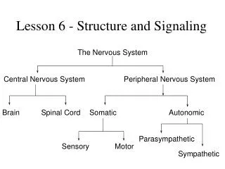
The Nervous System. Lesson 6 - Structure and Signaling. Central Nervous System. Peripheral Nervous System. Brain. Spinal Cord. Somatic. Autonomic. Parasympathetic. Sensory. Motor. Sympathetic. Nervous System. Afferent (sensory) Neurons – Carry impulses toward the CNS

Lesson 6 - Structure and Signaling
E N D
Presentation Transcript
The Nervous System Lesson 6 - Structure and Signaling Central Nervous System Peripheral Nervous System Brain Spinal Cord Somatic Autonomic Parasympathetic Sensory Motor Sympathetic
Nervous System • Afferent (sensory) Neurons – Carry impulses toward the CNS • Efferent (Motor) Neurons – Carry impulses from the CNS • Interneurons – conduct impulses within the spinal cord (between afferent and efferent) (Syn. Association, Internuncial) • Ganglia are small masses of nervous tissue located outside the brain and spinal cord.
Sensory Neurons • INPUT Fromsensory organs to the brain and spinal cord. Brain Sensory Neuron Spinal Cord Touch receptors in skin Somatosensory neuron - spinal Vision, hearing, taste and smell - cranial
Motor Neurons Brain Spinal Cord Motor Neuron • OUTPUTFrom the brain and spinal cord to muscles and glands Motor neurons in spinal cord
Interneurons • PROCESSING Relay information between other neurons Brain Spinal Cord Inter- Neurons Interneurons in brain
Divisions of the PNS • Somatic Nervous System (SNS) • Sensory neurons that convey information from sensory receptors in the head, body wall and limbs to the CNS • Motor neurons from the CNS that conduct impulses to the skeletal (voluntary) muscles only.
Divisions of the PNS • Autonomic Nervous System (ANS) • Sensory neurons convey information from receptors in the viscera (internal organs), to the CNS. • Motor neurons then convey information from the CNS to smooth muscle, cardiac muscle, glands, etc. • Motor functions in the ANS are not normally under conscious control; they are involuntary.
Neuron Resting Potential • Uneven concentrations of Na+ (outside) and K+ (inside) on either side of neuron membrane results in the inside of the neuron being 70 mV less positive than the outside • Text, pg. 419
A sodium-potassium pump maintains resting membrane potential after ions “leak” down their concentration gradient - 3 Na+ ions are actively pumped out while 2 K+ ions are pumped in.
ACTION POTENTIAL • Nerve signals are transmitted by action potentials that are abrupt, pulse-like changes in the membrane potential that last a few ten thousandths of a second. • Action potentials can be divided into three phases: the resting or polarized state, depolarization, and repolarization • The amplitude of an action potential is nearly constant and is not related to the size of the stimulus, so action potentials are all-or-nothing events.
Terminology • Synapse • Region at which neurons come nearly together to communicate. (neuron or effector organ) • Synaptic Cleft • Gap between neurons (at a synapse) • Impulses can not propagate across a cleft • Synaptic Vesicle • Packets of neurotransmitter in presynaptic neuron • Presynaptic Neuron • Neuron sending a signal (before the synapse) • Postsynaptic Neuron • Neuron receiving a signal (after the synapse)
Neurotransmitters 5 general criteria: 1) synthesized and released by neurons 2) released at the nerve terminal in a 'chemically identifiable' form 3) the chemical should reproduce the activity of the presynaptic neuron 4) can be blocked by competitive antagonist based on concentration 5) active mechanisms to stop the function of the neurotransmitter Classical transmitters are small molecules (often amino acid based) Non-classical transmitters can be peptides or even gasses
5 Steps of Neurotransmission 1) synthesis of the neurotransmitter precursors and enzymes should be in the correct place 2) storage of neurotransmitter OR precursor often stored in presynaptic vesicles
5 Steps of Neurotransmission 3) release of the neurotransmitter generally by vesicle fusion 4) binding to target receptor ionotropic receptors open ion channels metabotropic receptors modulate other signals
5 Steps of Neurotransmission 5) termination of the signal active termination caused by reuptake or chemical breakdown *For e.g. acetylcholine is broken down by . . . passive termination uses diffusion
“FLIGHT OR FIGHT RESPONSE” • Sympathetic component prepares body for stress [neurotransmitter used=norepinephrine] • Diverts blood from internal organs to skeletal muscles, heart & brain • Parasympathetic brings things back to normal [neurotransmitter used=acetylcholine] • Work in conjunction/opposition to each other • Ex. “on” / “off” switches
You come across a bear on your walk to school…what happens? • Sympathetic nervous system does what? • Increases heart rate • Increases breathing rate • Dilates bronchioles • Dilates pupils • Inhibits digestion