Download
1 / 12
140 likes | 422 Vues
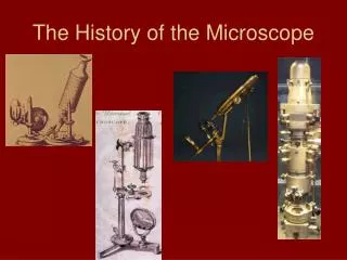
Microscope History and Development (2). Field of view and Magnification Check and go over yesterday’s HW p 140-1. Early Microscopes - Anton Van Leeuwenhoek . The father of microscopy, Anton Van Leeuwenhoek of Holland ( 1632-1723 ).

E N D
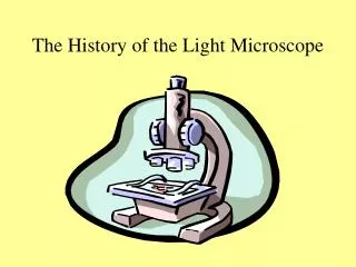
Microscope History and Development(2) Field of view and Magnification Check and go over yesterday’s HW p 140-1
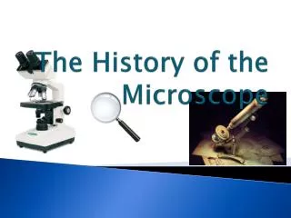
Early Microscopes - Anton Van Leeuwenhoek • The father of microscopy, Anton Van Leeuwenhoek of Holland (1632-1723). • Anton Van Leeuwenhoek was the first to see and describe bacteria (1674), yeast plants, the teeming life in a drop of water, and the circulation of blood corpuscles in capillaries.
Robert Hooke • In 1665, the English physicist Robert Hooke looked at a sliver of cork through a microscope lens and noticed some "pores" or "cells" in it. • Hooke was the first person to use the word "cell" to identify microscopic structures when he was describing cork.
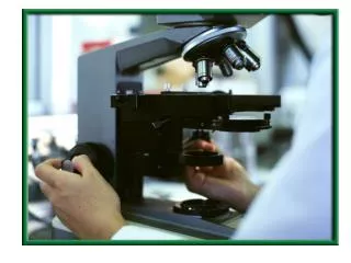
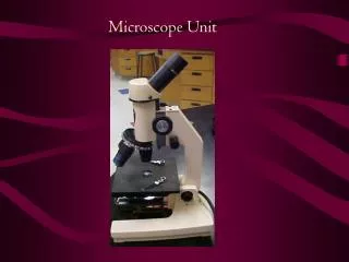
Compound Light Microscopes • Uses light • Has two lenses • Magnification limited to 2000x (400x at LHHS)
Transmission Electron Microscope (TEM) • Uses beams of electrons • Magnification of 2 000 000x • Has two limitations: • Good only for thin specimens • Only dead cells can be observed
Scanning Electron Microscope (SEM) • Electrons are reflected from the surface of the specimen • Produces a 3-d image • Good for the thicker specimens • Lacks the magnification and resolution of the transmission electron microscope
Magnification Magnification = Objective lens X Ocular lens (4x, 10x, 40x) (10x)
Calculating the size of a specimen • binder
Calculating the size of a specimenExample under med. objective Object size = Size of field of view Number of objects across field of view Object size = 1.72 mm 14 Object size = 0.1 mm