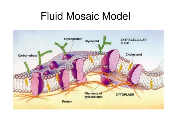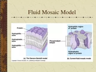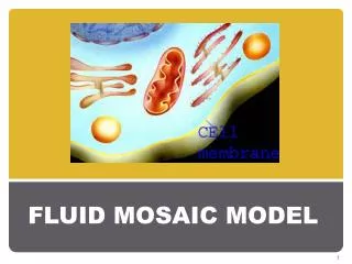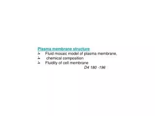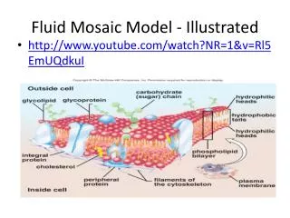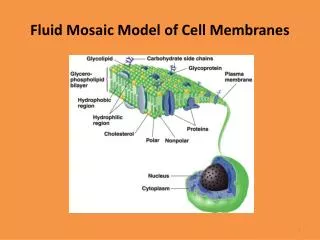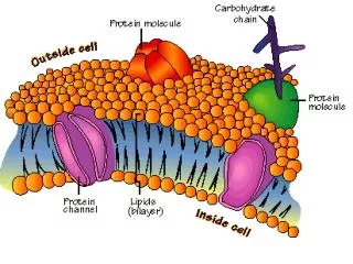
Fluid Mosaic Model
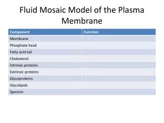
Fluid Mosaic Model. Figure 3.3. Functions of Membrane Proteins. Transport Enzymatic activity Receptors for signal transduction. Figure 3.4.1. Functions of Membrane Proteins. Intercellular adhesion Cell-cell recognition Attachment to cytoskeleton and extracellular matrix. Figure 3.4.2.

Fluid Mosaic Model
E N D
Presentation Transcript
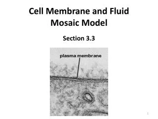
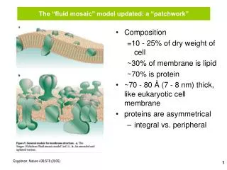
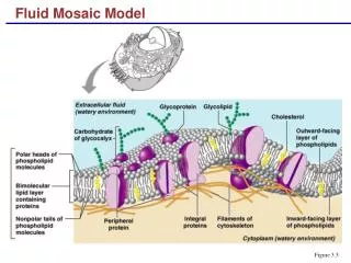
Fluid Mosaic Model Figure 3.3
Functions of Membrane Proteins • Transport • Enzymatic activity • Receptors for signal transduction Figure 3.4.1
Functions of Membrane Proteins • Intercellular adhesion • Cell-cell recognition • Attachment to cytoskeleton and extracellular matrix Figure 3.4.2
Membrane Junctions • Tight junction – impermeable junction that encircles the cell • Desmosome – anchoring junction scattered along the sides of cells • Gap junction – a nexus that allows chemical substances to pass between cells
Membrane Junctions: Tight Junction Figure 3.5a
Membrane Junctions: Desmosome Figure 3.5b
Membrane Junctions: Gap Junction Figure 3.5c
Diffusion Through the Plasma Membrane Figure 3.7
Passive Membrane Transport: Diffusion • Facilitated diffusion • Transport of glucose, amino acids, and ions • Transported substances bind carrier proteins or pass through protein channels
Active Transport • Uses ATP to move solutes across a membrane • Requires carrier proteins
Sodium-Potassium Pump Extracellular fluid K+ is released and Na+ sites are ready to bind Na+ again; the cycle repeats. 6 Binding of cytoplasmic Na+ to the pump protein stimulates phosphorylation by ATP. 1 Cytoplasm Phosphorylation causes the protein to change its shape. 2 Concentration gradients of K+ and Na+ The shape change expels Na+ to the outside, and extracellular K+ binds. 3 Loss of phosphate restores the original conformation of the pump protein. 5 K+ binding triggers release of the phosphate group. 4 Figure 3.10
Types of Active Transport • Primary active transport – hydrolysis of ATP phosphorylates the transport protein causing conformational change • Secondary active transport – use of an exchange pump (such as the Na+-K+ pump) indirectly to drive the transport of other solutes
Types of Active Transport • Symport system – two substances are moved across a membrane in the same direction • Antiport system – two substances are moved across a membrane in opposite directions (more common)
Types of Active Transport Figure 3.11
Vesicular Transport • Transport of large particles and macromolecules across plasma membranes • Endocytosis – enables large particles and macromolecules to enter the cell • Exocytosis – moves substance from the cell interior to the extracellular space
Vesicular Transport • Transcytosis – moving substances into, across, and then out of a cell • Vesicular trafficking – moving substances from one area in the cell to another • Phagocytosis – pseudopods engulf solids and bring them into the cell’s interior
Vesicular Transport • Fluid-phase endocytosis – the plasma membrane infolds, bringing extracellular fluid and solutes into the interior of the cell • Receptor-mediated endocytosis – clathrin-coated pits provide the main route for endocytosis and transcytosis • Non-clathrin-coated vesicles – caveolae that are platforms for a variety of signaling molecules
Clathrin-Mediated Endocytosis Figure 3.13
Exocytosis Figure 3.12a