LYMPHADENOPATHY
LYMPHADENOPATHY. By. Prof. Dr. Sameh Shamaa. Def.: It is an abnormal increase in size, or altered consistency of L.N . & is a clinical manifestation of regional or systemic disease & serve as an excellent clue to the underlying pathology and aetiology .

LYMPHADENOPATHY
E N D
Presentation Transcript
LYMPHADENOPATHY By Prof. Dr. Sameh Shamaa
Def.: It is an abnormal increase in size, or altered consistency of L.N. & is a clinical manifestation of regional or systemic disease & serve as an excellent clue to the underlying pathology and aetiology.
Causes of Localised Lymphadenopathy • 1- L.N Draining a Septic Focus: • * cervical : tonsilitis, scarlet fever, scalp infection. • * periauricular: otitis media. • * axillary : infections of fingers • * inguinal : infections of lower extremities. • * popliteal : infections of heel. 2-Carcinomatous L.N. Draining a Malignant Tumour: * hilar and scalene: bronchus. * virchow’s: stomach * cervical: thyroid, tongue, parotid.
Causes of LocalisedLymphadenopathy(2) 3- Systemic Infections * Viruses: a- Viral hepatitis Rt. supraclavecular L.N b- L.G.V. --groin (ing LN) c- German measles (cervical LN) * Bacteria: a- Plague b- T.B * Spirochetes : (Iry $ draining chancer) a- penis inguinal L.N. b- lips submandibular L.N. c-nipple axillary L.N. * Protozoa a -Filarial infectious-----inuguinal L.N. N.B Generalized L.N. may start as localized L.N. as in Hodgkin’s disease
Causes of Generalised Lymphadenopathy I- Infectious * Viruses: a-Infectious mononucleosis b-Cytomegalo virus (C.M.V.) * Bacteria: a- brucellosis b- T .B. *Spirochetes: (2ry $) *Protozoa a-kalaazar b-toxoplasmosis.
Causes of GeneralisedLymphadenopathy(2) • 2- Leukaemias: especially chronic lymphocytic leukamia (C.L.L.) • 3- Lymphomas:a- Hodgkin’s disease (H.D.) • b-Non- Hodgkin’s lymphoma (N.H.L) • 4- Collagenosis: a-rheumatoid artheritis. • b- Felty’s syndrome. • c-Still's disease. • d- D.L.E. • 5-Allergy: e.g., - Serum sickness. • 6- Sarcoidosis • 7- Lipoidosis • 8-Miscellaneous
Characters of L.N. Enlargement in Some Diseases 1- Streptococcal infection of tonsils: *Uni or Bilateral * Tender & unmatted *Usually submandibular but may extend to lower cervical group. 2- Scarlet Fever * Sore throat. * marked enlargement of submandibularL.N. *Other cervical L.N. (bilateral, tender, discrete, suppuration is common). 3-Diphtheria *Enlarged submandibularL.N. usually bilateral, tender, not matted.
4-German Measle: *OccipitaIL.N. enlargement are nearly always present, closely resembles that of infectious mononucleosis. 5-Infectious Mononucleosis: * Sore throat, Fever, sometimes headache, myalgia. * Bilateral L.N. enlargement, firm, discrete, mobile. * Appear first in posterior cervical area, adjacent to cervical spines, few days later , submandibular L.N. will be enlarged * Palatal petechiae often, are present * Mild splenomegally in 50% of cases *Lymphocytosisin 75% of cases with some atypical lymphocytes.
6- T.B.: * The chiefly affected group is upper cervical group, generalized L.N. enlargement is exceptional. * Unilateral or Bilateral. * Often firm, matted, painful, may become adherent to skin or deep structures. * Cystic areas may occur due to caseation and later on cold abscess formation. *Overlying skin may break down giving T.B. ulcers or sinuses.
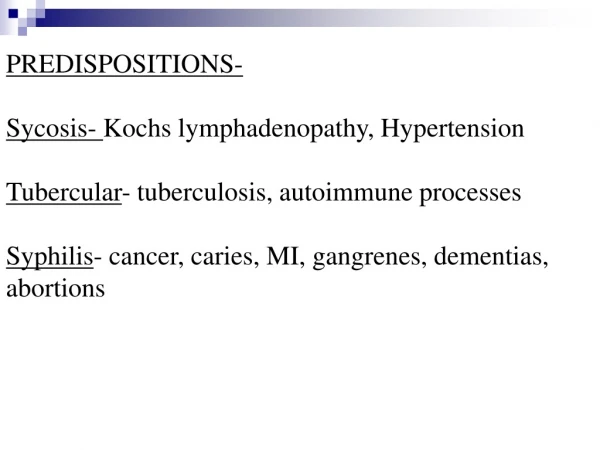
7-Syphilis: *Iry $:- L.N draining a chancre -Rocky hard, uni Or bilateral, not tender. *2ry $:- -Generalized L.N. enlargement especially posterior triangle of the neck or epitrochleargp (slightly enlarged, shotty, discrete, painless).
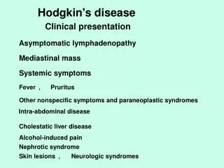
8- LYMPHOMATOUS L. N: *May be associated with constitutional symptoms.(anorexia, fever, weight loss, sweating, ….. etc). * Pel Ebstein fever: may be observed in H.D., it is a period of fever lasting for few days or weeks alternating with longer or shorter apyrexial periods . * L.N. usually discrete at start & not tender (but may become tender during febrile periods). * L.N. may increase in size during pyrexial periods and decrease in size during apyrexial periods
a-H.D.: • * may be confined to one group at first esp. lower cervical group then later on generalized L.N. enlargement. • Glands are: • a- moderately enlarged, not tender. • b- Firm, rubbery in consistency. • c- Discrete, mobile however as a result of later extension outside the capsule glands become matted or fixed • b-N.H.L: • *Also the cervical group is firstly affected • *Rapid rate of growth results in large number of variable sized nodes which are hard in consistency, tend to become fused and fixed to deep structures & may give pressure manifestations.
9- LEUKAEMIC L. N: *May be associated with general manifestations (fever, malaise, anorexia, headache, Hemorhagictendency) a- Acute Leukaemia: *Late, slightly or moderately enlarged *Soft, discrete esp. cervical L.N. due to oral sepsis *May be tender bone. b-C.L.L: * May affect cervica1 L.N. but mostly all superficial L.N. are enlarged. *The glands usually are (firm, not tender, not matted, usually moderately enlarged, but in advanced stages may be markedly enlarged) c-C.M.L.: *Rare to be manifested by L.N. enlargement.
10- CARCINOMATOUS L.N.: *Firm, but some times hard. *A stoney hard nodes fixed to underlying tissues are nearly always neoplastic in nature, however the reverse is not true. *Carcinomatous L.N. may be freely mobile
Clinical Approach Presentation: * Swelling * Constitutional symptoms (fever, sweating, loss of wt., pruritis) * Pressure symptoms: -Mediastinal syndrome - Pressure on veins →oedema - Pressure on nerves → pain Age: * T.B.: usually in children and young adults. * H.D. : any age including childhood, but its highest incidence ( ) (20-40 ys) * N.H.L.: usually at middle age and late life. * Acute Leuk.: any age, but highest in first 6 years of life.
History: * of infections, drugs, or vaccinations. Distribution: * Localized or generalized. * Single or multiple groups affected. Characters mentioned before. Other Signs: *Fever:Leuk., H.D., N.H.L., PelEbsteinfever. *Jaundice :H.D.,Chronicleuk. (due to hepatic infiltration → pressure on bile duct) *Eye: in leuk. (infections, sub conj. Hge., exophthalmos.) * Mouth, Tonsils, Parotid, Gums.
* Skin: • -pruritis, esp. H.D., N.H.L., Leuk. • - Rash in L.G. • - skin nodules in C.L.L., NHL. • - herpes zoster in H.D. • * Genitalia: chancre, chancroid, ulcer, gonorrhea • *Mediastinum & Chest : mediastinal syndrome, pleural effusion. • * Tenderness of sternum : in C.M.L. • *Bone tenderness. & Pathological fractures : in H.D. & N.H.L. • *Abdomen: • - ascitis and masses • - liver: • i- acute leuk : late & slightly enlarged. • ii-C.M.L. : firm & smooth • iii- C.L.L. : enlarged liver • - spleen : huge in C.M.L. & may be friction rub
* N.B.: in H.D.: 2/3 moderately enlarged spleen. I /3 moderately enlarged liver. in Sarcoidosis :hepatosplenomegaly in 1/3 of cases * Limbs : bone aches, swelling, joint affection. * C.N.S: - esp. M.D. & N.H.L. - brain & spinal cord : Hge, meningeal infiltration, pressure manifest. - peripheral nerves: pain, parathesia. -mediastinum : Horner's syndrome, or vocal cords
Investigations *For cases of genera1ised lymphadenopathy or local L.N. enlargement without local cases: (1) Complete clinical examinations. (2) C.B.P. & E.S.R. (3) Serological tests for infections mononucleosis,T.B. toxoplasmosis, $. (4) Plain chest X ray. (5) Biopsy. (6) Bone marrow aspiration if leuk. is suspected from C.B.P. * Biopsy should be done for enlarged L.N. of more than one month duration and not responding to usual ttt.
* According to L.N. biopsy ?: if: - +ve→ management. - -ve (single reactive hyperplasia) →follow up & if persist repeat biopsy two months later. Isolated Mediastinal L.N. Enlargement occurs in: - H.D. & N.H.L. - T.B. & Sarcoidosis. - Cancer lung or oesophagus. Isolated Abdominal L.N. Enlargement occurs in: - H.D & N.H.L - Metastasis.
Staging of Lymphoma A- Clinical staging: 1-Detaild history esp. in systemic symptoms. 2-Clinical examination including the Waldeyer's ring & areas of bone metastasis. 3-Adequate surgical biopsy 4- Routine lab. tests (C.B.P. & E.S.R & liver kidney function tests & Serum Uric acid.) 5- Plain chest X ray (P.A & Lat. view) 6- Bilateral lower extremities lymphangiography. 7- Radiological examinations (G.I.T., Gastroscopy if + veWaldeyer's ring) 8- Abdominal Ultrasonography or C.T. Scan.
B- Pahological staging: 1- Bone narrow biopsy. 2- Staging labarotomy. (only indicated in H.D with clinical stage I & II, if theraputic decision will depend on the identification of occult abdominal involvement) .
The Ann Arbor Staging Classification:- Stage I* Involvement of single L.N. region (I) * Or single extra nodal organ or site (IE*) Stage II. *involvement of two or L.N region on the same side of the diaphragm (II). *or. localised involvement of an extranodal organ or site will one or more L.N. regions on the same side of the diaphragm (IIE*).
The Ann Arbor Staging Classification:- Stage III III: involvement of L.N. regions on both sides of diaphragm. IIIs: may be also accompanied by splenic enlargement IIIE* : or by localized involvement of an extranodal site. IIISE* : or both. Stage IV * Diffuse or disseminated involvement of one or more extranodal organs with or without associated L.N. involvement.
NB. • * Any of them is further subdivided into: • A : without systemic symptoms. • B : with systemic symptoms • * (E) means very limited extra- lymphatic disease (both in site & extent) subjected to define ttt by radiotherapy. e.g: • i: L.N. + adjacent bone • ii: Ant. mediastinum + sternal invasion. • iii: Mediastinal L.N. + adjacent lung tissues.