Download
1 / 27
380 likes | 2.62k Vues
Approach to A child with cervical lymphadenopathy. Professor Pushpa Raj Sharma Department of Child Health Institute of Medicine. Location of enlarged nodes. The horizontal nodes are positioned at the junction of the head with the neck .

E N D
Approach to A child with cervical lymphadenopathy Professor Pushpa Raj Sharma Department of Child Health Institute of Medicine
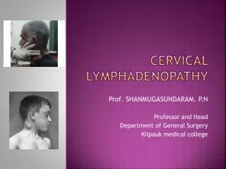
Location of enlarged nodes The horizontal nodes are positioned at the junction of the head with the neck The vertical nodes drain the deep structures of the head and neck
Infective Tender (not in tuberculosis) Acute onset Evidence of infection in drainage area Soft/fluctuant Local Non-infective Non tender Chronic onset Evidence of systemic manifestation Firm/hard Generalized Approach to a child with lymphadenopathy
Common infectious causes:Bacterial • Group A streptococcus • Mycobacteria: typical and atypical • Anaerobic bacteria • Diphtheria • Brucellosis • Actinomycetes • Gram –ve enterios
Common infectious causes:Viral • Epstein-Barr virus • Herpes simplex • Measles • Mumps • Coxsackie • Adenovirus • HIV • Rubella
Common infectious causes:Fungal / *Parasitic • Aspergillosis • Candida • Cryptococcus • Histoplasmosis • Coccidioidomycosis • Sporotrichosis • Blastomycosis • Toxoplasmosis*
Common Non Infectious Causes:Malignancy • Hodgkin’s/Non-Hodgkin’s Lymphoma • Leukaemia • Neuroblastoma • Thyroid tumours • Metastatic • Rhabdomyosarcoma
Common Other Causes: • Kawasaki Disease • Immunodeficiency diseases • Autoimmune disease (SLE, Still’s disease) • Castleman disease • Histiocytosis X • Serum sickness • Sarcoidosis
Mimicking Lymphadenopathy: • Branchial cleft cyst • Cystic hygroma • Thyroglossal duct cyst • Epidermoid cyst • Sternocleidomastoid tumor
10 year old; Male from Ramechap Swelling in the neck 5 months Fever for one month Weight: 15 Kg; Height: 113 cms Physical Exam – Multiple lymph nodes in the neck; vertical and horizontal; non tender; mobile; other: unremarkable CASE PRESENTATION
This case • Non tender • Chronic onset • No evidence of fungal disease • No evidence of autoimmune disease Possible diagnosis: • Tubercular • Malignancy • Sarcoidosis
Had a routine CXR Blood: WBC: 7,000/cmm; N: 72%; L: 28%; Hb: 8.4gm%. Mediastinal mass: a. Malignancy b. Tubercular c. Sarcoidosis Investigations
Mediastinal Mass • Mediastinum- Region between the pleural sacs • Tumors arise from anterior, middle & posterior compartments
Extent of Mediastinum • Anterior - sternum anteriorly to pericardium & brachiocephalic vessels posteriorly • Middle - between the anterior & posterior compartments • Posterior - pericardium & trachea anteriorly to vertebral column posteriorly
Anterior Mediastinum: Contents • Thymus • Anterior mediastinal lymph nodes • Internal mammary A & V • Pericardial fat
Middle Mediastinum: Contents • Heart & Pericardium, ascending aorta & arch of aorta, vena cavae, brachiocephalic A &V , • phrenic nerve • trachea, main stem bronchi & contiguous lymph nodes • Pulmonary A & V
Posterior Mediastinum: Contents • Descending thoracic aorta • Esophagus • Thoracic duct • Azygos & hemiazygos vein • Posterior group of mediastinal nodes • Sympathetic trunk & intercostal nerves
Origins of Mediastinal Mass • Developmental • Neoplastic • Infectious • Traumatic • Cardiovascular disorders
Anterior Mediastinal Masses: • Thymoma • Teratoma • Thyromegaly • Lymphoma • Lipoma, Fibroma - rare
Middle Mediastinal Masses: • Aneurysms - aorta, innominate artery, enlarged pulmonary artery • Lymphadenopathy secondary to carcinoma / metastasis / granulomatosis • Cysts - enteric, bronchogenic, pleuropericardial • Dilated azygos, hemiazygos veins • Hernia of Foramen of Morgagni
Posterior Mediastinal Masses: • Neurogenic tumors • Meningo-myelocele, meningocele • Esophageal - tumor, cyst, diverticula • Hiatus hernia • Hernia of Foramen of Bochdalek • Thoracic spine disease, • Extramedullary hematopoiesis
DIAGNOSTIC APPROACH • Imaging - CT, MRI, Radionuclide study, • Tissue sampling - Mediastinoscopy, Thoracoscopy, Needle aspiration, Open Biopsy • Barium study for hernia, achalasia, diverticula • I-131 for intrathoracic goiter
DIAGNOSTIC APPROACH • Mediastinoscopy or anterior mediastinotomy can definitively diagnose anterior & middle mediastinal masses • Video assisted thoracoscopy plays an important role in diagnosis
TREATMENT & PROGNOSIS • Dictated by the etio-pathology of the mass
This case • Nospecific- no pressure effect of mass sorrounding structures • Chronic onset with fever and loss of weight • mass detected on CXR • Physical findings : cervical lymphadenopathy; fever; loss of weight. • 50% mediastinal masses are malignant in children
Histopathology of the lymph node showing caseating necrosis and Langhans’ type giant cells (arrow).
This case: • Non tender cervical lymph node • Apyrexial • CXR: mass in the anterior mediastinum • Lungs normal • Biopsy of cervical lymphnode suggestive of tuberculosis