Plasma Membrane Structure and Function
Explore the composition and function of the plasma membrane, including phospholipid bilayers, proteins, and fluidity. Learn about membrane proteins and their various roles in cell function. Discover the mechanisms of membrane transport and diffusion.

Plasma Membrane Structure and Function
E N D
Presentation Transcript
Membrane Structure and Function Chapter 7 A. P. Biology
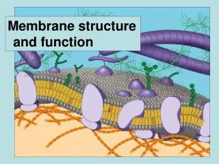
Plasma Membrane • Also called the plasmalemma. • Plasma Membrane = Phospholipid Bilayer + Transmembrane Proteins + Supporting Fibers + Glycoproteins and Glycolipids
WATER Hydrophilic head Hydrophobic tail WATER Figure 7.2 Scientists studying the plasma membrane • Reasoned that it must be a phospholipid bilayer
Phospholipid Bilayer • Glycerol + 2 Fatty Acids + Phosphorylated Alcohol = Phospholipid • Hydrophilic or Polar Region = Phosphate • Hydrophobic or Nonpolar region = Fatty Acids
The Davson-Danielli sandwich model of membrane structure • Stated that the membrane was made up of a phospholipid bilayer sandwiched between two protein layers. • Was supported by electron microscope pictures of membranes
Hydrophobic region of protein Phospholipid bilayer Figure 7.3 Hydrophobic region of protein In 1972, Singer and Nicolson • Proposed that membrane proteins are dispersed and individually inserted into the phospholipid bilayer
APPLICATION A cell membrane can be split into its two layers, revealing the ultrastructure of the membrane’s interior. TECHNIQUE Extracellular layer Proteins Knife Plasma membrane Cytoplasmic layer These SEMs show membrane proteins (the “bumps”) in the two layers, demonstrating that proteins are embedded in the phospholipid bilayer. RESULTS Freeze-fracture studies of the plasma membrane • Supported the fluid mosaic model of membrane structure A cell is frozen and fractured with a knife. The fracture plane often follows the hydrophobic interior of a membrane, splitting the phospholipid bilayer into two separated layers. The membrane proteins go wholly with one of the layers. Extracellular layer Cytoplasmic layer Figure 7.4
Lipid Bilayer • Nonpolar interior prevents passage of water-soluble, polar compounds. • Only very small, uncharged molecules like O2 and H2O can enter through the lipid bilayer. • Also, allows nonpolar compounds to freely enter.
Lateral movement (~107 times per second) Flip-flop (~ once per month) (a) Movement of phospholipids Figure 7.5 A The Fluidity of Membranes • Phospholipids in the plasma membrane • Can move within the bilayer
Fluid Viscous Unsaturated hydrocarbon tails with kinks Saturated hydro- Carbon tails (b) Membrane fluidity Figure 7.5 B The type of hydrocarbon tails in phospholipids • Affects the fluidity of the plasma membrane
Cholesterol Figure 7.5 (c) Cholesterol within the animal cell membrane The steroid cholesterol • Has different effects on membrane fluidity at different temperatures
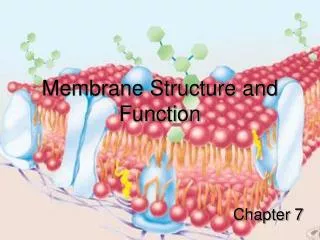
Glycoprotein Carbohydrate Glycolipid EXTRACELLULAR SIDE OF MEMBRANE Microfilaments of cytoskeleton Peripheral protein Cholesterol Integral protein CYTOPLASMIC SIDE OF MEMBRANE Figure 7.7 Membrane Proteins and Their Functions • A membrane • Is a collage of different proteins embedded in the fluid matrix of the lipid bilayer Fibers of extracellular matrix (ECM)
Lipid Bilayer is Fluid • Fluid = Moving, Dynamic. • Each lipid can rotate, move laterally • Fluidity depends on temperature and type of fatty acid used. • Unsaturated fatty acids are more fluid. • Fluid Mosaic Model
Transmembrane Proteins (Integral Proteins) • Part of the protein that extends through the bilayer is nonpolar (several nonpolar amino acids in this region). • Usually is an alpha helix or beta barrel. • Used to anchor protein in the membrane. • Beta-barrels = form a pore and are called a porin protein.
N-terminus C-terminus CYTOPLASMIC SIDE a Helix Figure 7.8 Integral proteins • Penetrate the hydrophobic core of the lipid bilayer • Are often transmembrane proteins, completely spanning the membrane Extracellular Side EXTRACELLULAR SIDE
(a) Transport ATP (b) Enzymes Enzymatic activity (c) Signal Signal transduction Receptor Figure 7.9 • An overview of six major functions of membrane proteins
(d) Cell-cell recognition Glyco- protein (e) Intercellular joining (f) Attachment to the cytoskeleton and extracellular matrix (ECM). Figure 7.9
Transmembrane Proteins • Channels = passive transport of molecules across membrane. • Carriers = transport of molecules against the gradient. • Receptors = transmit information into the cell. • Cell Adhesion Proteins = connect cells to each other. • Cytoskeleton Attachment Proteins = to attach actin.
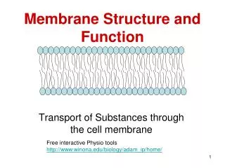
Movement Across the Membrane • Diffusion = random motion of molecules that causes a net movement from areas of high concentration to areas of low concentration. • Osmosis = diffusion of water across a selectively permeable membrane.
Factors that Affect the Direction of Diffusion • The concentration gradient; High Low. • Temperature; High heat Low Heat. • Pressure; High Pressure Low Pressure
Factors that Affect the Rate of Diffusion • The steepness of the gradient. • The molecular weight of the solute.
Concentrations • Osmotic concentrations = concentrations of all solutes in a solution. • If unequal concentrations… Hyperosmotic = solution with the higher solute concentration. Hypoosmotic = solution with the lower solute concentration. Isosmotic = solutions with the same osmotic or solute concentration.
Crenate Plasmolyzes
Osmotic Pressure • If a cell’s cytoplasm is hyperosmotic to the extracellular fluid, then water diffuses into the cell and it swells. Pressure of the cytoplasm pushing out against the membrane- hydrostatic pressure. • Osmotic pressure is the pressure needed to stop the osmotic movement of water across a membrane.
How do living things maintain osmotic balance? • Some oceanic eukaryotes adjust internal [solutes]- they are isosmotic. • Animals – circulate an isosmotic fluid around their cells. Must constantly monitor the fluid’s [solute] Ex. Humans secrete albumin into the plasma to match the body cells.
Protozoa- are hyperosmotic, so use extrusion to remove excess water; may have special organelles-contractile vacuoles. • Plants- are hyperosmotic, but do not circulate an isosmotic solution; are usually under osmotic pressure- turgor pressure-presses the plasma membrane against the cell wall.
(b) Plant cell. Plant cells are turgid (firm) and generally healthiest in a hypotonic environ- ment, where the uptake of water is eventually balanced by the elastic wall pushing back on the cell. H2O H2O H2O H2O Turgid (normal) Flaccid Plasmolyzed Figure 7.13 • Water balance in cells with walls
Bulk Movement through Membranes • Endocytosis- the cytoskeleton extends the membrane outward toward food particles. Bulk transport into cell. • Extended membrane encircles the particle, fuses with itself, and contracts. • Forms a vesicle around particle.
Three Kinds of Endocytosis • Phagocytosis - “cell eating”- large, amounts of organic material;white blood cells and protists. • Pinocytosis- “cell drinking”- liquid material brought into cell; mammalian ova and follicle cells. • Receptor-mediated Endocytosis- use receptors in the membrane for specific transport into cell.
Receptor-Mediated Endocytosis • Have indentations on the plasma membrane. • Indentations = are clathrin-coated pits. • Pits have receptor proteins on the extracellular side = trigger • When receptor binds to target molecule, clathrin proteins on the cytoplasmic side begins endocytosis. • Forms a clathrin -coated vesicle. • Very specific.
Exocytosis • The release of material from vesicles at the cell surface. • Examples: protists using a contractile vacuole to release water, gland cells secreting hormones, neurons releasing neurotransmitters.