MEMBRANE STRUCTURE AND FUNCTION
MEMBRANE STRUCTURE AND FUNCTION. Yasir Waheed. Plasma membrane encloses the cell, defines its boundaries and maintains the essential difference between the cytosol and extracellular environment.

MEMBRANE STRUCTURE AND FUNCTION
E N D
Presentation Transcript
MEMBRANE STRUCTURE AND FUNCTION Yasir Waheed
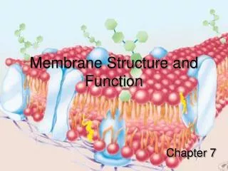
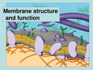
Plasma membrane encloses the cell, defines its boundaries and maintains the essential difference between the cytosol and extracellular environment. • Inside the eukaryotic cell , various membrane enclosed organelles (like mitochondria, Golgi bodies ) maintains the difference between each organelle and the cytosol. • Despite of small differences , all biological membranes have same structure: composed of a very thin film of lipid and protein molecules, held together by non covalent interactions. • Inside the plasma membrane, lipid molecules are arranged as a continuous double membrane of about 5nm thickness. • Lipid molecules serve as a impermeable barrier to the transport of water soluble molecules. • Protein molecules embedded in the membrane, have both structural and functional role.
THE LIPID BILAYER • Membrane Lipids are amphipathic molecules • Lipids are fatty acid molecules, constitute about 50% of the mass of most animal cell membranes, nearly all of the remainder being protein. • There are approximately 5 × 10⁶ lipid molecules in a 1 mm × 1 mm area of lipid bilayer or about 109 lipid molecules in the plasma membrane of a small animal cell. • All of the lipid molecules in cell membranes are amphipathic that is, they have a hydrophilic ("water-loving") or polar end and a hydrophobic ("water-fearing") or nonpolar end. • The most abundant membrane lipids are the phospholipids. These have a polar head group and two hydrophobic hydrocarbon tails. The tails are usually fatty acids, and they can differ in length (they normally contain between 14 and 24 carbon atoms). • Hydrophobic tails are buried in the interior of the membrane while the hydrophilic heads are exposed to water.
Four major phospholipids predominate in the plasma membrane of many mammalian cells: phosphatidylcholine, phosphatidylethanolamine, phosphatidylserine, and sphingomyelin. Only phosphatidylserine carries a net negative charge, the other three are electrically neutral, carrying one positive and one negative charge. Together these four phospholipids constitute more than half the mass of lipid in most membranes. In Apoptosis, the cell displays phosphatidyserine on its surface, recognized by the apoptotic machinery , results in cell death.
Phosphatidylcholine and sphingomyelin are present on outer monolayer of the membrane, while Phosphatidyl ethanol amine and Phosphatidylserine are located in the inner monolyaer of the membrane. When animal cells undergo programmed cell death, or apoptosis , phosphatidylserine, which is normally confined to the cytosolic monolayer of the plasma membrane lipid bilayer, rapidly translocates to the extracellular monolayer. The phosphatidylserine exposed on the cell surface serves as a signal to induce neighboring cells, such as macrophages, to phagocytose the dead cell and digest it.
Glycolipids These are the lipid molecules with sugar residues and are present on the cytosolic face of the membrane. Addition of sugars on lipid molecules takes place in the lumen of the Golgi bodies. Figure 10-16. Glycolipid molecules. (A) Galactocerebroside is called a neutral glycolipid because the sugar that forms its head group is uncharged. (B) A ganglioside always contains one or more negatively charged sialic acid residues (also called Nacetylneuraminic acid, or NANA), whose structure is shown in (C).
Membrane Proteins Basic structure of the bilayer is formed by the lipid molecules, proteins forms their specific functions, vary from cell to cell. A typical plasma membrane contains proteins about 50% by mass. Plasmamembrane contains seven different types of proteins, some of them are the transmembrane , rest of them are attached with membrane on either intracellular or extracellular face. Some proteins are peripheral proteins and some of them are integral proteins. Some of the transmembrane proteins attached with prenly group (fatty acid) with the plasma membrane.
Figure 10-17. Various ways in which membrane proteins associate with the lipid bilayer. Most trans-membrane proteins are thought to extend across the bilayer as (1) a single a helix, (2) as multiple a helices, or (3) as a rolled-up beta sheet . Some of these "single-pass" and "multipass" proteins have a covalently attached fatty acid chain inserted in the cytosolic lipid monolayer (1). Other membrane proteins are exposed at only one side of the membrane. (4) Some of these are anchored to the cytosolic surface by an amphipathic alpha helix that partitions into the cytosolic monolayer of the lipid bilayer through the hydrophobic face of the helix. (5) Others are attached to the bilayer solely by a covalently attached lipid chain either a fatty acid chain or a prenyl group in the cytosolic monolayer or, (6) via an oligosaccharide linker, to phosphatidylinositol in the noncytosolic monolayer. (7, 8) Finally, many proteins are attached to the membrane only by noncovalent interactions with other membrane proteins.
In Most Transmembrane Proteins the Polypeptide Chain Crosses the Lipid Bilayer in an alpha-Helical Conformation A segment of a transmembrane polypeptide chain crossing the lipid bilayer as an a helix. Only the alpha backbone of the polypeptide chain is shown, with the hydrophobic amino acids in green and yellow.
A single-pass transmembrane protein. The polypeptide chain traverses the lipid bilayer as alpha helix and that the oligosaccharide chains and disulfide bonds are all on the noncytosolic surface of the membrane. The sulfhydryl groups in the cytosolic domain of the protein do not normally form disulfide bonds because the reducing environment in the cytosol maintains these groups in their reduced (-SH) form.
Figure 10-21. Beta barrels formed from different numbers of beta strands. (1) The E. coli OmpA protein (8 b strands), which serves as a receptor for a bacterial virus. (2) The E. coli OMPLA protein (12 b strands), is a lipase that hydrolyses lipid molecules. The amino acids that catalyze the enzymatic reaction (shown in red) protrude from the outside surface of the barrel. A porin from the bacterium Rhodobacter capsulatus, which forms water-filled pores across the outer membrane (16 b strands). The diameter of the channel is restricted by loops (shown in blue) that protrude into the channel. (4) The E. coli Fep A protein (22 b strands), which transports iron ions. The inside of the barrel is completely filled by a globular protein domain (shown in blue) that contains an iron-binding site. This domain is thought to change its conformation to transport the bound iron, but the molecular details of the changes are not known.
The recognition that biological membranes are two-dimensional fluids was a major advance in understanding membrane structure and function. It has become clear, however, that the picture of a membrane as a lipid sea in which all proteins float freely is greatly oversimplified. Many cells have ways of confining membrane proteins to specific domains in a continuous lipid bilayer. In epithelial cells, such as those that line the gut or the tubules of the kidney, certain plasma membrane enzymes and transport proteins are confined to the apical surface of the cells, whereas others are confined to the basal and lateral surfaces. The specific distribution of the proteins in a plasma membrane is crucial for the function of the cell.
Figure 10-39. An experiment demonstrating the mixing of plasma membrane proteins on mouse-human hybrid cells. The mouse and human proteins are initially confined to their own halves of the newly formed heterocaryon plasma membrane, but they intermix with time. The two antibodies used to visualize the proteins can be distinguished in a fluorescence microscope because fluorescein is green whereas rhodamine is red.
Figure 10-45. Simplified diagram of the cell coat (glycocalyx). The cell coat is made up of the oligosaccharide side chains of glycolipids and integral membrane glycoproteins and the polysaccharide chains on integral membrane proteoglycans. Note that all of the carbohydrate is on the noncytosolic surface of the membrane.
The hydrophobic interior of the lipid bilayer of cell membranes serves as a barrier to the passage of most polar molecules. • This barrier function is crucially important because it allows the cell to maintain concentrations of solutes in its cytosol that are different from those in the extracellular fluid and in each of the intracellular membrane enclosed compartments. • To make use of this barrier, however, cells have had to evolve ways of transferring specific water-soluble molecules across their membranes in order to ingest essential nutrients, excrete metabolic waste products, and regulate intracellular ion concentrations. • The transport of inorganic ions and small water soluble organic molecules across the lipid bilayer is achieved by specialized transmembrane proteins, each of which is responsible for the transfer of a specific ion, molecule, or group of closely related ions or molecules. • Cells can also transfer macromolecules and even large particles across their membranes, but the mechanisms involved in most of these cases are different from those used for transferring small molecules. • The importance of membrane transport is indicated by the large number of genes in all organisms that code for transport proteins, which make up between 15 and 30% of the membrane proteins in all cells. Some specialized mammalian cells devote up to two-thirds of their total metabolic energy consumption to membrane transport processes.
* The cell must contain equal quantities of positive and negative charges (that is, be electrically neutral). Thus, in addition to Cl-, the cell contains many other anions not listed in this table; in fact, most cellular constituents are negatively charged (HCO3-, PO43-, proteins, nucleic acids, metabolites carrying phosphate and carboxyl groups, etc.). The concentrations of Ca2+ and Mg2+ given are for the free ions. There is a total of about 20 mM Mg2+ and 1-2 mM Ca2+ in cells, but this is mostly bound to proteins and other substances and, for Ca2+ , stored within various organelles.
Figure 11-1. The relative permeability of a synthetic lipid bilayer to different classes of molecules. The smaller the molecule and, moreimportantly, the less strongly it associates with water, the more rapidly the molecule diffuses across the bilayer.
There Are Two Main Classes of Membrane Transport Proteins: Carriers and Channels • Carrier proteins (also called carriers, permeases, or transporters) bind the specific solute to be transported and undergo a series of conformational changes to transfer the bound solute across the membrane. • Channel proteins, in contrast, interact with the solute to be transported much more weakly. They form aqueous pores that extend across the lipid bilayer; when these pores are open, they allow specific solutes (usually inorganic ions of appropriate size and charge) to pass through them and thereby cross the membrane. Transport through channel proteins occurs at a much faster rate than transport mediated by carrier proteins.
Figure 11-3. Carrier proteins and channel proteins. (A) A carrier protein alternates between two conformations, so that the solute-binding site is sequentially accessible on one side of the bilayer and then on the other. (B) In contrast, a channel protein forms a water-filled pore across the bilayer through which specific solutes can diffuse.
Figure 11-4. Passive and active transport compared. (A) Passive transport down an electrochemical gradient occurs spontaneously, either by simple diffusion through the lipid bilayer or by facilitated diffusion through channels and passive carriers. By contrast, active transport requires an input of metabolic energy and is always mediated by carriers that harvest metabolic energy to pump the solute against its electrochemical gradient.
IONOPHORES • Ionophores are small hydrophobic molecules that dissolve in lipid bilayers and increase their permeability to specific inorganic ions. • They are widely used by cell biologists as tools to increase the ion permeability of membranes in studies on synthetic bilayers, cells, or cell organelles. • There are two classes of ionophoresmobile ion carriers and channel formers. • Both types operate by shielding the charge of the transported ion so that it can penetrate the hydrophobic interior of the lipid bilayer. • Ionophores are not coupled to energy sources, they permit the net movement of ions only down their electrochemical gradients. • Valinomycin is an example of a mobile ion carrier. It is a ring-shaped polymer that transports K+ down its electrochemical gradient by picking up K+ on one side of the membrane, diffusing across the bilayer, and releasing K+ on the other side.
Carrier Proteins and Active Membrane Transport • Carrier protein has one or more specific binding sites for its solute (substrate). It transfers the solute across the lipid bilayer by undergoing reversible conformational changes that alternately expose the solute-binding site first on one side of the membrane and then on the other. • Coupled carriers couple the uphill transport of one solute across the membrane to the downhill transport of another. • 2. ATP-driven pumps couple uphill transport to the hydrolysis of ATP. • 3. Light-driven pumps, which are found mainly in bacterial cells, couple uphill transport to an input of energy from light, as with bacterio-rhodopsin
Figure 11-8. Three ways of driving active transport. The actively transported molecule is shown in yellow, and the energy source is shown in red.
Figure 11-9. Three types of carrier-mediated transport. This schematic diagram shows carrier proteins functioning as uniporters, symporters, and antiporters.
One way in which a glucose carrier can be driven by a Na+ gradient. The carrier oscillates between two alternate states, A and B. In the A state, the protein is open to the aextracellular space; in the B state, it is open to the cytosol. Binding of Na+ and glucose is cooperative that is, the binding of either ligand induces a conformational change that greatly increases the protein's affinity for the other ligand. Since the Na+ concentration is much higher in the extracellular space than in the cytosol, glucose is more likely to bind to the carrier in the A state. Therefore, both Na+ and glucose enter the cell (via an A to B transition) much more often than they leave it (via B to A transition). The overall result is the net transport of both Na+ and glucose into the cell. Because the binding is cooperative, if one of the two solutes is missing, the other fails to bind to the carrier. Thus, the carrier undergoes a conformational switch between the two states only if both solutes or neither are bound.
Na + K + Pump • The concentration of K+ is typically 10 to 20 times higher inside cells than outside, whereas the reverse is true of Na+. These concentration differences are maintained by a Na + K + pump, or Na + pump, found in the plasma membrane of virtually all animal cells. • The pump operates as an antiporter, actively pumping Na+ out of the cell against its steep electrochemical gradient and pumping K+ in. Because the pump hydrolyzes ATP to pump Na+ out and K+ in, it is also known as a Na + , K +ATPase. • An essential characteristic of the Na+ K+ pump is that the transport cycle depends on autophosphorylation of the protein. The terminal phosphate group of ATP is transferred to an aspartic acid residue of the pump and is subsequently removed.
The Na+ K+pump. This carrier protein actively pumps Na+ out of and K+ into a cell against their electrochemical gradients. For every molecule of ATP hydrolyzed inside the cell, three Na+ are pumped out and two K+ are pumped in. The specific inhibitor ouabain and K+ compete for the same site on the extracellular side of the pump.
(1) The binding of Na+ and (2) the subsequent phosphorylation by ATP of the cytoplasmic face of the pump induce the protein to undergo a conformational change that (3) transfers the Na+ across the membrane and releases it on the outside. (4) Then, the binding of K+ on the extracellular surface and (5) the subsequent dephosphorylation return the protein to its original conformation, which (6) transfers the K+ across the membrane and releases it into the cytosol. The Na+ dependent phosphorylation and the K+ dependent dephosphorylation of the protein cause the conformational transitions to occur in an orderly manner, enabling the protein to do useful work.
Protein function • Plasma membrane proteins serve diverse functions including: • Transport • Enzymatic activity • Signal transduction • Intercellular joining • Cell-cell recognition • Attachment to the cytoskeleton and extracellular matrix