S 3
S 3. S 3.1 Positive ionization low energy CID fragmentation spectrum of the singly phosphorylated peptide pSLKPDTENQESSVK of Rec 10

S 3
E N D
Presentation Transcript
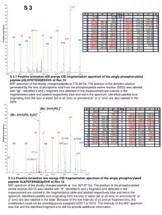
S 3 S 3.1 Positive ionization low energy CID fragmentation spectrum of the singly phosphorylated peptide pSLKPDTENQESSVK of Rec 10 MS3 spectrum of the doubly charged peptide at 772.38 Da. The position of the dehydro-alanine generated by the loss of phosphoric acid from the phosphorylated serine residue (S503) was labeled with “@”. Identified b and y fragment ions detected in the measurement are colored in the fragmentation table and labeled respectively blue and red in the spectrum. Identified satellite ions originating from the loss of water (b0 or y0 ions) or ammonia (b* or y* ions) are also labeled in the table. S 3.2 Positive ionization low energy CID fragmentation spectrum of the singly phosphorylated peptide SLKPDTENQESpSVK of Rec 10 MS2 spectrum of the doubly charged peptide at m/z 821.67 Da. The position of the phosphorylated serine residue (S513) was labeled with “#”. Identified b and y fragment ions detected in the measurement are colored in the fragmentation table and labeled respectively blue and red in the spectrum. Identified satellite ions originating from the loss of water (b0 or y0 ions) or ammonia (b* or y* ions) are also labeled in the table. Because of the low intensity of y3 and y4 fragment ions, the modification could not be unambiguously assigned toS511 or S512. The intensity of the MS3 spectrum was low and the identified fragment ions did not provide additional information.
S 3.3 Positive ionization low energy CID fragmentation spectrum of the singly phosphorylated peptide AKSNVNLQYpSPK of Rec 10 MS3 spectrum of the doubly charged peptide at m/z 665.94 Da. The position of the dehydro-alanine generated by the loss of phosphoric acid from the phosphorylated serine (S529) residue was labeled with “@”. Identified b and y fragment ions detected in the measurement are colored in the fragmentation table and labeled respectively blue and red in the spectrum. Identified satellite ions originating from the loss of water (b0 or y0 ions) or ammonia (b* or y* ions) are also labeled in the table. S 3.4 Positive ionization low energy CID fragmentation spectrum of the singly phosphorylated peptide NRpSIIQIK of Rec 10 MS3 spectrum of the doubly charged peptide at m/z 477.46 Da. The position of the dehydro-alanine generated by the loss of phosphoric acid from the phosphorylated serine (S356) residue was labeled with “@”. The MS3 spectrum showed an intensive fragment ion at m/z 468.96 Da. This can be interpreted as the loss of ammonia from the arginine residue after the loss of phosphoric acid from the phosphoserine residue. Identified b and y fragment ions detected in the measurement are colored in the fragmentation table and labeled respectively blue and red in the spectrum. Identified satellite ions originating from the loss of water (b0 or y0 ions) or ammonia (b* or y* ions) are also labeled in the table.
S 3.5 Positive ionization low energy CID fragmentation spectrum of the singly phosphorylated peptide SNVNLQYpSPK of Rec 10 MS3 spectrum of the doubly charged peptide at m/z 566.32 Da. The position of the dehydro-alanine generated by the loss of phosphoric acid from the phosphorylated serine residue (S529) was labeled with “@”. The loss of water from the unphosphorylated serine residue generated an intensive fragment ion, which is also labeled. Identified b and y fragment ions detected in the measurement are colored in the fragmentation table and labeled respectively blue and red in the spectrum. Identified satellite ions originating from the loss of water (b0 or y0 ions) or ammonia (b* or y* ions) are also labeled in the table. S 3.6 Positive ionization low energy CID fragmentation spectrum of the singly phosphorylated peptide DSLpSADDYAYDTK of Rec 10 MS3 spectrum of the doubly charged peptide at m/z 723.23 Da. The position of the dehydro-alanine generated by the loss of phosphoric acid from the phosphorylated serine (S424) residue was labeled with “@”. Identified b and y fragment ions detected in the measurement are colored in the fragmentation table and labeled respectively blue and red in the spectrum. Identified satellite ions originating from the loss of water (b0 or y0 ions) or ammonia (b* or y* ions) are also labeled in the table.
S 3.7 Positive ionization low energy CID fragmentation spectrum of the singly phosphorylated peptide LLPAIIVpSPK of Rec 10 MS2 spectrum of the doubly charged peptide at m/z 566.12 Da. The fragment ion generated by the loss of phosphoric acid from the phosphoserine residue (S347) was labeled in the spectrum. The intensity of this fragment was not among the 3 most intensive ones due to the strong fragmentation at the proline residues, thus no MS3 spectrum was generated. Identified b and y fragment ions detected in the measurement are colored in the fragmentation table and labeled respectively blue and red in the spectrum. The position of the phosphoserine residue was labeled with “#”. Identified satellite ions originating from the loss of water (b0 or y0 ions) or ammonia (y* ions) are also labeled in the table. S 3.8 Positive ionization low energy CID fragmentation spectrum of the singly phosphorylated peptide DENVINQTGPAKKpTPVQRRK of Rec 10 MS3 spectrum of the triply charged peptide at m/z 754.78 Da. The position of the dehydro-aminobutyric acid generated by the loss of phosphoric acid from the phosphorylated threonine (T482) residue was labeled with “@”. Identified b and y fragment ions detected in the measurement are colored in the fragmentation table and labeled respectively blue and red in the spectrum. This peptide was generated by AspN digest and contained several basic amino acids. Because of this reason several doubly or triply charged fragment ions could be detected.
S 3.9 Positive ionization low energy CID fragmentation spectrum of the ubiquitinylated peptide VIKEFSK of Rec 10 MS2 spectrum of the doubly charged peptide at m/z 482.8 Da. Identified b and y fragment ions detected in the measurement are colored in the fragmentation table and labeled respectively blue and red in the spectrum. The position of the modified residue (K757) was labeled with “^”. Identified satellite ions originating from the loss of water (b0 or y0 ions) or ammonia (y* ions) are also labeled in the table. S 3.10 Positive ionization low energy CID fragmentation spectrum of the ubiquitinylated peptide SSVWKELLKE of Rec10 MS2 spectrum of the doubly charged peptide at m/z Da. Identified b and y fragment ions detected in the measurement are colored in the fragmentation table and labeled respectively blue and red in the spectrum. The position of the modified residue (K604) was labeled with “^”. Identified satellite ions originating from the loss of water (b0 or y0 ions) or ammonia (y* ions) are also labeled in the table.
S 3.11 Positive ionization low energy CID fragmentation spectrum of the ubiquitinylated peptide NEAYNPSSKSATIDGLQR of Rec 10 MS2 spectrum of the triply charged peptide at m/z 689.57 Da. Identified b and y fragment ions detected in the measurement are colored in the fragmentation table and labeled respectively blue and red in the spectrum. The position of the modified residue (K712) was labeled with “^”. Identified satellite ions originating from the loss of water (b0 or y0 ions) or ammonia (y* ions) are also labeled in the table.
S 3.12 Positive ionization low energy CID fragmentation spectrum of the ubiquitinylated peptide LIFAGKQLEDGR of Ubi2 MS2 spectrum of the doubly charged peptide at m/z 731.42 Da. Identified b and y fragment ions detected in the measurement are colored in the fragmentation table and labeled respectively blue and red in the spectrum. The position of the modified residue (K48) was labeled with “^”. Identified satellite ions originating from the loss of water (b0 or y0 ions) or ammonia (y* ions) are also labeled in the table.


![CP = E[ s 2 , s 5 , s 1 , s 3 , s 2 ’ , s 3 ’ , s 4 , s 1 ’ , s 4 ’ , s 5 ’ ] S[] I[]](https://cdn3.slideserve.com/6546826/slide1-dt.jpg)