Dermatology
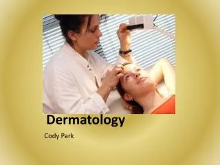
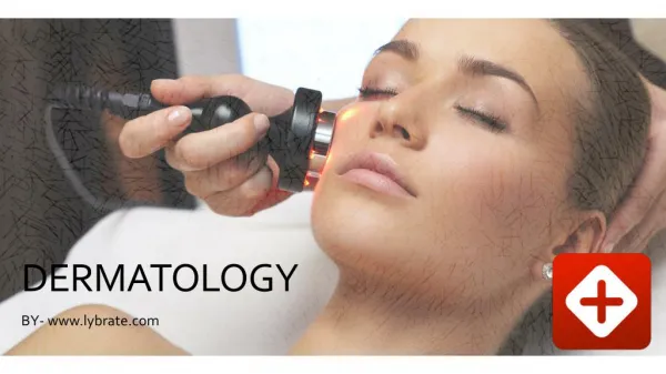
Dermatology. Vickie Mickey,CT. Dermatology. Study of skin, diseases, and treatment Electrologist encounter the presence of skin disorders and diseases Electrologist must be able to identify dermatology disorders in order to know when to treat or not treat a skin disorder

Dermatology
E N D
Presentation Transcript
Dermatology Vickie Mickey,CT
Dermatology • Study of skin, diseases, and treatment • Electrologist encounter the presence of skin disorders and diseases • Electrologist must be able to identify dermatology disorders in order to know when to treat or not treat a skin disorder • Certain skin disorders are contraindicated
Lesion • Lesions are a structural change in the tissue caused by trauma or disease • Three types of lesions: 1. Primary 2. Secondary 3. Tertiary or vascular
Primary Lesions • First to recognize • Flat or non-palpable • Skin color changes may occur • Elevation can occur • Solid mass
Macule • Small discolored spot or patch on the surface of the skin, flat or level with skin • A freckle, flat moles, liver spot (chloasma) or leukoderma (loss of pigment) • Pink or red inflammatory macules – indicative of typhoid, typhus, secondary syphilis, or allergenic drug reaction • Blue Macules in public regions indicate crab lice
Papule • Small elevated pimple • Contains no fluid but may develop pus
Common Papular Eruptions • Pityriasis rosea – self-limited disease of truck, eruptions at time barely evident • Mucocutaneous syphilis – papules tend to be juicy, easily molded, found in mouth and genital region • Psoriasis – chronic inflammatory disease of skin, lesions are round, dry papules, covered with coarse slivery scales
Common Papule Eruptions • Warts – verruca • Seborrheic keratois – flat, brown, greasy feeling papules, waxy texture. Non-malignant • Actinic keratosis – hard, horny papules, results of sun and wind exposure. May become malignant • Senile keratosis– hard horney papules, old age spots • Molluscum contagiosum – virus infection, pin-head sized to pea-sized, wax like papules of a yellowish white or pinkish color
Common Papule • Acne – different stages • Drug eruptions – may occur in many forms • Scratch a localized reaction to rubbing, scratching
Wheal • Itchy, swollen lesion that last only a few hours • Hive or insect bite • Capable of returning
Nodule • Solid lump, larger than a papule, projects above the surface or may lie below the skin • Varies in size and color • Of medical significance if nodule appears in more than one area
Nodules • Nevis – birthmark • Lipomas – fatty growth cells • Fibromas – benign tumor composed of whorls of white fibrous connective tissue. May occur in single or in random areas, not indicative of underlying disease
Tumor • Abnormal swelling or enlargement that can be malignant or benign • Fluidless lesions
Vesicle/Bulla • Raised lesion containing fluid derived from serum of blood • Water fluid collects within epidermis or between epidermis and dermis, ranging color from clear to pink • Bulla is larger than a Vesicle
Vesicle/Bulla • Herpes Simplex – virus • Contact Dermatitis – inflammation resulting from irritation of certain substances • Sun burn – (second degree) • Blister – skin irritation
Pustule • Small round, round, raised area of inflamed skin filled with pus • Common types: acne • Furuncles and carbuncles – contain infection • Impetigo – highly contagious inflammatory disease • Eczema • Acute fungal infection of feet and hands • Smallpox, chickenpox, vaccination reaction, herpes
Secondary Lesion • Secondary lesions are primary lesion that have progressed to later stage of disease • Secondary lesions usually characterized by material buildup on the skin and includes:
Scale • Scale is dry and greasy accumulation of epidermal flakes • Silvery, stratified scales of psoriasis • Seborrhea dermatitis – yellow greasy scales caused by excess secretion of sebaceous glands
Crust • Crust/Scab – dry accumulation of serum and pus, mixed with epidermal material • Lymph crust – honey colored scab not oozing, evidence of skin healing • Lymph crust – can appear after electrolysis • Pus Crust – a dirty gray or greenish-yellow crust indicative of infection • Blood crust is reddish or black crust from dried blood, indicative of disease of blood
Excoriation • Skin sore produced from scratching or abrasion
Fissure • Crack in skin that penetrates the dermis
Ulcer • Open skin lesion or mucous membrane that is accompanied by pus and loss of skin depth
Keloid • Skin or film that forms over a wound • Keloids rarely form on face • Keloids appear more in skin of color
Scare • Specialized growth of tissue that forms during healing of wound or skin condition
Common Skin Diseases • Herpes Simplex – an acute inflammatory infection of the skin, occurring as swollen vesicle or group • Appears around mouth or nostrils • DO NOT TREAT AN AREAS With Herpes!
Impetigo • An acute inflammatory and highly contagious disease • Usually found in children • Characterized by honey colored crust that have a “stuck on appearance! • DO NOT TREAT THIS SKIN CONDITION
Psoriasis • Reddish patches covered by silvery scales • Tiny bleeding points may appear under scales • Not infectious • Electrologist can work on this skin involved with however, never within the patches
Dermatitis • Irritation with vesicular eruptions • External on skin • Wait until skin has resolved before treating with electrolysis
Parasitic Disease of the Skin • Parasites both animal or vegetable can affect skin • Very Contagious • Refer to physician • DO NOT TREAT
Scabies • Very contagious • Itching skin caused by infestation by itch mite • Mites burrow in hands or feet • Skin reacts with eruption of papules on forearms, underarms, waist, inner thigh, buttocks, and ankles • If disease is not treated it may manifest into boils and ulcers • DO NOT TREAT
Pediculosis • Head lice – Pediculois Capitis • Transmitted from person to person via infected personal care items • Nit surround hair and passes up out of pore with growing hair • Body Louse – Pediculosis Corporis • Causes intense itching • Clothing contains nits
Disease of Sebaceous Glands • Blackheads – comedomes – hardened plug of sebaceous material • Appears black as the sebum oxidizes and turns black • Can block mouth of follicle • Can become inflamed
Acne • Chronic inflammation of the philosebaceous unit • Common on face, back and chest • May present as papules or nodules • Can appear as pustules • Cysts may appear • Acne begins as a blockage of the follicular canal, develops through leakage of sebum into the dermis
Acne • Once the dermis becomes irritated it can develop into a red pustule or pimple • Prevalent in adolescence during heightened androgenic activity stimulates sebaceous activity • Infected follicles can be treated with electrolysis
Milia • Whitehead caused by retention cyst of entrapped sebum beneath the epidermis • Not in follicle pore • Appears on face, neck, shoulders and chest areas • Not usually associated with hair follicle
Sebaceous Cyst • Subcutaneous tumor of the sebaceous glands • Composed of sebum • Ranges in size from small pea to an orange • Releasing sebum will not eliminate cyst • Needs to be removed by physician
Seborrhea • Oily condition of nose, forehead, and scalp • Result of over production of sebaceous glands • Can accompany itching and burning
Asteatosis • Dry, scaly skin deficient of sebum
Disorder of Sudoriferous Glands • Hyperidrosis – excessive perspiration • Caused by stimulates or mental depression and exhaustions • Great condition to treat with laser on underarms • Bromidrosis – foul smelling perspiration • Appears in armpits, or feet • Can be caused by drugs, foods, or rare disease • Result of apocrine sweat glands
Disorders of Sudoriferous Glands • Anidrosis – lack of perspiration caused by fever or disease • Miliaria or Heat Rash – an acute inflammatory disorder of the sudoriferous glands • Lesions consists of vesicles and papules
Pigmentation Irregularities • Tan – Protective layer of skin • Sunlight barrier for skin • Prevents excessive production of Vitamin D
Freckles & Lentigines • Yellowish or brown spots occurring on hands, face, and other exposed body parts • Result of sun exposure
Chloasma • Yellow, brown or black deposits in skin • Patches occurring in various shapes and spots • Appears a mask of pregnancy – can result from birth control pills • If patient displays in area to be treated with electrolysis , do test spots prior to treatment as it may increase with treatment • Laser hair removal often removes it
Stains • Abnormal patches of brown coloration • Round or irregular shapes
Nevus/ Birthmark • Colored malformation of the skin • Often congenital
Leucoderma • White patches of skin or all over body • Albinism – a congenital absences of pigment in skin, hair, eyes • Extremely sensitive to sun • Can be localized • Not a good candidate for laser! • Vitiligo – loss of pigment, occurring mostly in bands, often on hands and patches in the skin • Do not treat with laser!
Hypertrophies • Usually and enlargement of skin due to multiplication of cells • Occurs for many reasons • Keratoma (Callus) – hard thickened patches of skin due to hypertrophy of horny layer • Caused by irritation , friction, or pressure
Moles • Small congenital macule • Ranges in color from pale tan to brown to bluish black • True malignant moles is melanoma • Black, blue-black or slate-colored • Lacks hair • Mole that is normal for many years can suddenly change, increase in size, increase in depth, pigmentation • Crust can form and may bleed • Inflammatory ring may appear around it
Moles • When in doubt don’t treat a mole • Have checked by physician prior to treatment • Malpractice insurance may not cover if not checked prior to treatment • Get release to treat mole
Post Treatment Disorders • Over treatment can cause eschars (scabs) • Unsanitary practices by patient can result in infection • Examine skin disorder when confronted • Usually small areas will be related to the electrolysis treatment, one or two follicles • Good aftercare instruction very important to patient