Download
1 / 8
80 likes | 167 Vues
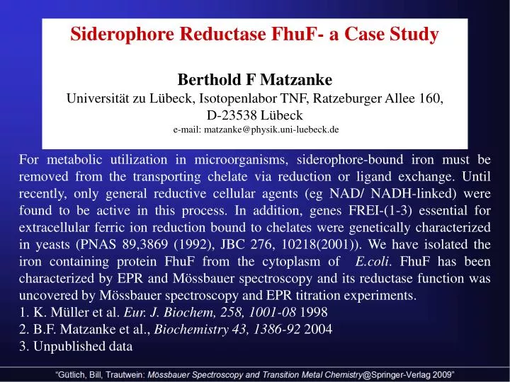
Siderophore Reductase FhuF- a Case Study Berthold F Matzanke Universität zu Lübeck, Isotopenlabor TNF, Ratzeburger Allee 160, D-23538 Lübeck e-mail: matzanke@physik.uni-luebeck.de.

E N D
Siderophore Reductase FhuF- a Case Study Berthold F Matzanke Universität zu Lübeck, Isotopenlabor TNF, Ratzeburger Allee 160, D-23538 Lübeck e-mail: matzanke@physik.uni-luebeck.de For metabolic utilization in microorganisms, siderophore-bound iron must be removed from the transporting chelate via reduction or ligand exchange. Until recently, only general reductive cellular agents (eg NAD/ NADH-linked) were found to be active in this process. In addition, genes FREI-(1-3) essential for extracellular ferric ion reduction bound to chelates were genetically characterized in yeasts(PNAS 89,3869 (1992), JBC 276, 10218(2001)). We have isolated the iron containing protein FhuF from the cytoplasm of E.coli. FhuF has been characterized by EPR and Mössbauer spectroscopy and its reductase function was uncovered by Mössbauer spectroscopy and EPR titration experiments. 1. K. Müller et al. Eur. J. Biochem, 258, 1001-08 1998 2. B.F. Matzanke et al., Biochemistry 43, 1386-92 2004 3. Unpublished data
FhuF- Protein of E. coli: Cells were grown in 57Fe-rich medium and the protein was isolated by chromatography.The Mössbauer spectra (Fig.1) display features of an -cys2[2Fe-2S]cys2-motif(S=1/2-system, parameters see table 1). EPR of mutants uncovered a hitherto unknown bis-cystein terminal iron-binding motif of the protein chain (data not shown): -cys-cys-(aa)10-cys-(aa)2-cys- (Fig. 2). Molecular modelling confirms that a bis-cystein-axial position is stable. However, the chelate ring exhibits strain.
Nuclear Hamiltonian used for the simulation of the Mössbauer spectra Q: nuclear quadrupole moment of excited nuclear state; Vzz: main component of the electric-field gradient (efg) tensor; = (Vxx - Vyy) / Vzz ; asymmetry parameter of efg. A: hyperfine coupling tensor; gN : nuclear g-factor. Do spectroscopic parameters reflect the strain caused by the bis-cystein axial ligand? Mössbauer spectra of a) the as-isolated protein b) of the dithionite reduced sample measured at 4.2 K in a field of 20 mT perpendicular to the -beam. The solid lines correspond to simulations based on the spin Hamiltonian formalism (from top to bottom): 1) as-isolated protein, 2) reduced cluster ferrous ion site, 3) reduced cluster ferric ion site. The reduced sample contains a portion of the as-isolated compound because the reduction was not complete
Mössbauer spectrum of dithionite reduced FhuF measured at 4.2 K in a field of 5.34 T perpendicular to the -beam. The solid lines correspond to spin Hamiltonian simulations of 1) the as-isolated contribu-tion, 2) the Fe2+-site and 3) the Fe3+-site of the reduced cluster (top to bottom) Mössbauer spectrum of oxidized FhuF measured at 4.2 K in a field of 7T parallel to the -beam. For the simulation the follow-ing parameters were used: =0.29 mms-1, EQ = 0.47 mms-1, S= 0, = 0.25 mms-1
The spectra of the reduced protein are fit best with an axially symmetric EFG tensor for the ferrous ion. The magnitude of EQ(Fe2+) compares well with other compounds of this class (2.7-3.6 mms-1). The same holds for (0.54-0.67 mms-1). However, the positive sign of EQ is solely shared with ferrochelatase. The spectra fit best with a rhombic ferrous A-tensor. Surprisingly, Azz is small-est in magnitude which is, again, in line with ferrochelatase and in contrast to other proteins of this class. The ferric ion site of the reduced cluster is simula-ted best with an axially symmetric EFG- and a cubic A-tensor. The quadrupole splitting differs from the majority of 2Fe-2S clusters (0.59-0.68 mm s-1) and groups together with murine ferrochelatase (1.2 mms-1) and Desulfovibrio gigas aldehyde oxidoreductase (1.0 mms-1). EQ (0.47 mms-1) of the as-isola-ted protein is the lowest of all [2Fe-2S]-proteins(0.6-0.7mms-1) analyzed so far. Oxidized protein at 4.3K, small velocity range: slight asymmetry, best fit with 2 ferric iron sites the iron sites are not exactly equivalent site1: = 0,281(1) EQ= 0.574(22) = 0.25, 50% site 2: = 0,287(2) EQ= 0.341(4) = 0.26, 50%
Summary: The Mössbauer parameters of the Fe3+-site are affected probably reflecting terminal dicystein binding. The Mössbauer- and EPR- parameters differ from other 2Fe-2S proteins: This is attributed to distortions caused by dicystein. Most spectroscopic similarities are found for murine ferrochelatase. Conclusion: Spectroscopic and structural features of the metal cluster reflect the difference of the two iron sites in terminal cystein binding. Has FhuF a physiological Role? Based on EPR-redox titration experiments a redox potential of -31025 mV was determined. According to the Nernst equation : favorable redox range is 59 mV, i.e. for FhuF the range extends from -394 mV to -236 mV. For the ferric siderophores the range is as follows: ferrichrome: from -469 to - 331 mV, coprogen: from -513 mV to- 378 mV and for ferrioxamine B from -542 to - 394 mV. The redox potential of FhuF should allow reduction of ferrichrome, coprogen and perhaps of ferrioxamine B. This was checked by Mössbauer spectroscopy: The reduced 57Fe FhuF protein was mixed with 57Fe-ferrioxamine (equimolar ratio)
Summary The differences of the Mössbauer parameters of FhuF compared to other [2Fe-2S]-proteins reflect its unique terminal bis-cystein iron binding. In addition, FhuF functions as a siderophore reductase, in fact, capable of reducing ferrioxamine B.