Download
1 / 23
350 likes | 782 Vues
Localized surface plasmon resonance of optically coupled metal particles. Takumi Sannomiya*, Christian Hafner**, Janos Vörös*. * Laboratory of Biosensors and Bioelectronics, IBT ** Computational Optics Group, IFH ETH Zürich. Outline. Background LSPR of colloid particles, motivations

E N D
Localized surface plasmon resonance of optically coupled metal particles Takumi Sannomiya*, Christian Hafner**, Janos Vörös* * Laboratory of Biosensors and Bioelectronics, IBT ** Computational Optics Group, IFH ETH Zürich
Outline BackgroundLSPR of colloid particles, motivations Method- MMP Calculation - Experimental setup Results- inter-particle coupling- selective excitation of coupling mode- possibility of more sensitive adsorption detection Summary & Outlook
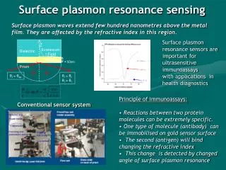
Resonance shift upon molecular binding Colloidal particle light Extinction l lr lr´ Molecule-adsorbed Localized Surface Plasmon Resonance(LSPR) for Sensors Adsorption Sensor Functionalize for certain molecules motivation: investigate adsorption of single colloid particle single molecule detection ?
Different colors illumination particle <<1/l Scattered light Objective Lens spectrometer How to take a spectrum of a single colloidal particle Dark field microscopy + spectrometer scattering spectrum Dark field image of 50nm Au colloids
Calculated Spectra Second peak appears Same as single particle Non-coupled mode Optically coupled gold colloid particles Electric field one mode for single particle coupled mode
Purpose To investigate optical property of coupled particles with the aid of numerical calculation • Dependence on Inter particle distance • Selective excitation of coupling mode • Possibility for better adsorption sensing?
Method y less order less parameters more order more parameters x z MMP Calculation (modeling) MMP: Multiple Multipole Program (MaX-1, Prof Ch.Hafner) Semi-analytical calculation proper location and order of multipoles Model medium boundary gold 3D multipole E S Symmetry plane y Calculated scattering intensity≡ Sz(0,0,500nm) 50nm z x εAu : P.B.Johnson et al; Phys.Rev. B v6.n12(1972)
Method Light source Dark Field Microscope polarizer CCD Camera sample Spectrometer Experimental setup - Spectrophotometer - HAL - Colloid sample - 50nm Au colloid particles were deposited on a glass slide by incubating the glass slide in colloid solution, rinsed and dried with nitrogen gas.
Result d =0.5nm =500nm =700nm =540nm =580nm =620nm Calculation: coupled mode
Result Calculation: non-coupled mode d =0.5nm =460nm =700nm =500nm =520nm =600nm
Result d E Separation Dependence(calculation) d = inter-particle separation - Coupled mode - Poynting Vector Map ( d = 0.5nm, l = 620nm ) • The second peak shifts to red as the distance decreases • Two particles behave independently when separated enough
Result d E Separation Dependence(calculation) - Non-coupled mode - Poynting Vector Map ( d = 1.0nm, l = 520nm ) No separation dependence Only the intensity is twice as high as single…
Result Experimental spectra of different particles Dark Field Image „Particle“ A „Particle“ B „Particle“ C by comparing with calculation
Result Coupling direction 90° 45° 135° polarization 0° 180° Experiment (selective excitation)
Result Coupling direction 90° 45° 135° polarization 0° 180° Experiment (selective excitation)
Result Experiment ( selective excitation) Intensity change by polarization 700nm> Longpass filtered Color: intensity polarization 2mm
Result Calculation (medium dependence) single particle coupled particle peak shift peak shift solution solution water water air air nwater = 1.333, nsolution = 1.370 separation : 0.75nm Peak of coupled mode is more sensitive to the surrounding refractive index sensitive to dimension sensitive to refractive index More sensitive adsorption detection !?
Result Experiment (medium dependence) air buffer solution single particle coupled particle Problem in air : water layer most sensitive part
Summary coupled mode…. • can be selectively excited by polarized light • is sensitive to the dimension and surrounding medium • can be a more sensitive adsorption detector Combination of MMP Calculation and experiment helps for better understanding and designing LSPR sensor. Outlook • Detect molecule adsorptions by coupled mode • Produce coupled particles by use of bio-molecules • Design new structures
Acknowledgements • Prof. Christian Hafner • Prof. Janos Vörös • LBB members Funding : ETH Zürich Thank you for the attention…
Result d =0.75nm d =0.75nm =500nm Calculation: three particles(symmetric separation) =900nm =580nm =620nm =760nm
Result Calculation: three particles(non-symmetric separation) d =1.5nm d =0.75nm =500nm =840nm =600nm =660nm =720nm
Result Calculation: three particles(symmetric separation) d =0.77nm d =0.75nm =900nm =500nm =560nm =700nm =740nm