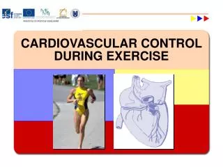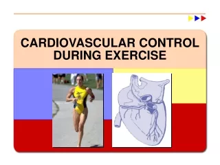
Cardiovascular Control During Exercise
Cardiovascular Control During Exercise. Learning Objectives. Review the structure and function of the heart, vascular system and blood. How the cardiovascular system responds to increased demands during exercise.
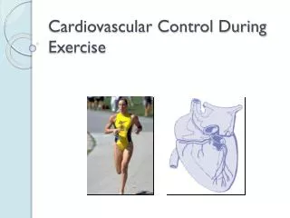

Cardiovascular Control During Exercise
E N D
Presentation Transcript
Learning Objectives • Review the structure and function of the heart, vascular system and blood. • How the cardiovascular system responds to increased demands during exercise. • Explore the role of the CV system in delivering oxygen and nutrients to active body tissue.
Major CV Function • Delivery of oxygen and other nutrients • Removal of CO2 and other metabolic waste • Transport of hormones • Thermoregulation • Maintenance of acid-base balance and overall body fluid balance • Immune function
Components of CV system • Heart (the pump) • Blood vessels (system of channels or tubes) • Blood (a fluid medium)
Heart To body From body To lungs From lungs
Myocardium- Cardiac Muscle • Myocardial thickness varies according to amount of stress placed on it • Cardiac muscle fibers interconnected by intercalated disks • Allows for rapid transmission of action potentials for uniform contractions • Myocardial fibers are homogenous and contain only one fiber type (similar to type I)
Left Ventricle • Most powerful of the four chambers • Must contract to pump blood through entire body (to the systemic circulation) • Higher the intensity of exercise, working muscles require more blood, increase need in LV to deliver blood to exercising muscles, therefore, LV will hypertrophy
Skeletal Muscle Heart Muscle Intercalated Discs
Cardiac muscle contraction occurs by “calcium-induced calcium release” • Calcium enters cell • Calcium released from SR • Primary blood supply to heart provided by right and left coronary arteries • Ability of myocardium to contract as a single unit depends on cardiac conduction system
Cardiac Conduction System • Cardiac muscle is able to generate own electrical signal-spontaneous rhythmicity • With no neural or hormonal stimulation, intrinsic HR is ~100 bpm • Four main components: • Sinoatrial (SA) node • Atrioventricular (AV) node • AV bundle (bundle of His) • Purkinje fibers (terminal branches of AV bundle)
SA Node • Group of specialized cardiac muscle fibers located in upper posterior wall of right atrium • Where impulse for normal heart contractions is initiated • Known as heart’s pacemaker because generates electrical impulse at ~100 bpm
AV Node • Conducts electrical impulse from atria to ventricles • Delayed by ~0.13 s as it passes through AV node and into AV bundle • Delay allows blood from atria to empty into ventricles to maximize ventricular filling before ventricles contract
AV bundle • Runs along ventricular septum and sends right and left bundle branches into both ventricles • Branches send impulse toward apex then outward • Purkinje Fibers • Transmit impulse 6x faster than through rest of cardiac conduction system
Extrinsic Control of The Heart • Parasympathetic nervous system (PNS) • Sympathetic nervous system • Endocrine system (hormones)
Parasympathetic Nervous System • Originates in medulla oblongata and reaches heart through vagus nerve • Vagus nerve carries impulses to SA and AV nodes • when stimulated, releases acetylcholine hyperpolarization of conduction cells decrease in HR • Predominates at rest: “vagal tone” • Able to decrease HR to 20-30 bpm
Sympathetic Nervous System • Increases HR and contraction force of the ventricles • Allows HR to increase to 250 bpm • Predominate during physical or emotional stress (when HR >100 bpm) • When exercise begins, HR first increases due to withdrawal of vagal tone, then later due to SNS
Endocrine System • Catecholamines: norepinephrine and epinephrine • Released from adrenal medulla • Stimulate heart and increase its rate and contractility • Triggered by sympathetic nervous system during times of stress • Actions prolong sympathetic response
Exercise Training Affects on HR • Normal RHR: 60-100 bpm • RHR can decrease to 35 bpm with training • Increase parasympathetic stimulation (vagal tone) and decrease sympathetic activity
Electrocardiogram (ECG) • Electrical activity of the heart can be recorded to monitor cardiac changes or diagnose potential cardiac problems • Electrical impulses generated in heart conducted through body fluids to skin • Three basic components: • The P wave • The QRS complex • The T wave
The P Wave • Represents atrial depolarization • Occurs when electrical impulse travels from SA node through atria to AV node
The QRS Complex • Represents ventricular depolarization • Occurs as impulse spreads from AV bundle to Purkinje fibers and through the ventricles
The T Wave • Represents ventricular repolarization • Atrial repolarization cannot be seen because it occurs during QRS complex
Cardiac Arrhythmias • Disturbances in the normal sequence of cardiac events can lead to an irregular heart beat-arrhythmia • Bradycardia • RHR lower than 60 bpm • Tachycardia • RHR >100 bpm • In both, rhythm is normal but rate is altered
Other Arrhythmias • Premature ventricular contractions (PVCs) • Feeling of skipped or extra beats • Relatively common • Result from impulses originating outside of SA node
Other Arrhythmias • Atrial flutter- atria contracts 200-400 bpm • Atrial fibrillation- atria contracts in rapid and uncoordinated manner • Both are more serious arrhythmias that cause ventricular filling problems
Other Arrhythmias • Ventricular tachycardia- ≥3 consecutive premature ventricular contractions • Very serious • Can lead to ventricular fibrillation-contraction of ventricular tissue is uncoordinated • little to no blood pumped out of heart • cause of most cardiac deaths
Cardiac Cycle • Includes all mechanical and electrical events that occur during one heartbeat • Consists of chambers that undergo diastole (relaxation phase) and systole (contraction phase) • Diastole-chambers fill with blood, ventricles contract and send blood into aorta and pulmonary veins
Cardiac Cycle • One cardiac spans the time between one systole to another • Systole (ventricular contraction) starts during the QRS complex and ends in the T wave • Diastole (ventricular relaxation) occurs during T wave and to next contraction • Heart spends more time in diastole (~2/3 of time) than in systole (~1/3 of time)
The Wiggers Diagram: Events of Cardiac Cycle for LV Function
Stroke Volume (SV=EDV-ESV) • Volume of blood pumped per beat (contraction) • End-diastolic volume (EDV)-volume of blood in ventricle before contraction • End-systolic volume (ESV)-volume of blood in ventricle after contraction • i.e.) SV= 100ml-40ml= 60ml
Ejection Fraction • Fraction of blood pumped out of left ventricle in relation to the amount of blood present before the contraction • EF=SV/EDV x 100 • i.e.) EF=60ml/100ml x 100 = 60%
Cardiac Output (Q) • Total volume of blood pumped by the ventricle per minute • Q=HR x SV • i.e.) Q= 60 beats/min x 70 ml/min = 4,200ml/min or 4.2L/min
Vascular System: Closed System • Arteries: transport blood away from heart to arterioles • Arterioles: site of greatest control of circulation by SNS (resistance vessels) • Capillaries: where exchange between blood and tissues occur • Venules • Veins: transport blood back to heart
Blood Pressure • Pressure exerted by blood on vessel walls • Systolic blood pressure (SBP)-highest pressure in the artery • Diastolic blood pressure (DBP)-lowest pressure in the artery
Blood Pressure • Mean arterial pressure (MAP): average pressure • MAP=2/3 DBP + 1/3 SBP • MAP=DBP + [0.333 x (SBP-DBP)] • i.e.) MAP=80 + [0.333 x (120-80)] = 93mmHg
General Hemodynamics • Blood flows in closed-system because of pressure gradient between arterial and venous sides • Pressure, Flow and Resistance • Blood flows from high pressure to low pressure • MAP in aorta = ~100 mmHg • MAP in right atrium = ~0 mmHg
General Hemodynamics • Blood flow is proportional to pressure difference across system and inversely proportional to resistance • Blood flow = ∆pressure/resistance • Regulation of blood flow to organs accomplished by vasoconstriction and vasodilation
Distribution of Blood • Varies depending on immediate needs of a specific tissue • Skeletal muscle receives ~15% of blood flow at rest and up to 80% during heavy endurance exercise • Changes in distribution in Q controlled by SNS mainly by arteriolar diameter
Intrinsic Control of Blood Flow • Ability of local tissues to vasodilate or vasoconstrict arterioles and alter regional blood flow depending on tissue need • Metabolic-increased O2 demand, decreases in other nutrients, increases in by-products (CO2, K+, H+, lactic acid) • Endothelium mediated vasodilation: NO
Intrinsic Control of Blood Flow • Myogenic contraction: • Muscle contracts in response to an increase in pressure • Relaxes in response to a decrease in pressure
Extrinsic Neural Control • Redistribution at the system or body level controlled by sympathetic nerves • Increase in sympathetic nerve activity muscle cells contractconstricts blood vesselsdecreases blood flow • Sympathetic nerves keep vessels moderately constricted (vasomotor tone) • Redirects blood flow from areas of low need to areas of high need
Distribution of Venous Blood Most of the blood volume is in the veins at rest, particularly in the viscera
Integrative Control of BP • Normally maintained by reflexes from ANS • Baroreceptors: pressure sensors in aortic arch and carotid arteries • Chemoreceptors • Mechanoreceptors
Return of Blood to the Heart • CV system requires mechanical assistance to overcome force of gravity for venous return • Three basic mechanisms: • Valves in the veins: enables unidirectional blood flow, prevents backflow and pooling of blood • The muscle pump • The respiratory pump