Download
1 / 64
660 likes | 1.34k Vues
Cardiovascular Control During Exercise. Learning Objective: To understand the functional anatomy of the CV system. “How is the system designed anatomically to function physiologically ?”. 1. Introduction to Cardiovascular System. Key Points: Pulmonary Circulation Circuit Low Pressure

E N D
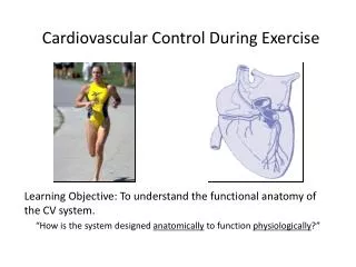
Cardiovascular Control During Exercise Learning Objective: To understand the functional anatomy of the CV system. “How is the system designed anatomically to function physiologically?”
1. Introduction to Cardiovascular System • Key Points: • Pulmonary Circulation Circuit • Low Pressure • O2 and CO2 Exchange (Hb Affinity) • Systemic Circulation Circuit • High Pressure • Overcome gravity/hydrostatic pressure • Bulk flow of blood to and from tissue • Key CV Components • Heart (the pump) • Blood Vessels (system of tubes) • Blood (a fluid medium) Figure from Marieb and Hoehn, 2010
Major CV Functions • O2 and Nutrient Delivery • CO2 and Metabolic Waste Removal • Transport (e.g. hormones) • Thermoregulation • Acid-base and Body Fluid Balance
Heart To body From body To lungs From lungs http://www.nhlbi.nih.gov/health/dci/Diseases/hhw/hhw_pumping.html
Myocardium- Cardiac Muscle • Adaptable: • Myocardial thickness will increase when “stressed” • Myocardial volume will increase with endurance training • Myocardial fibers are homogenous and contain only one fiber type (similar to type I)
Left Ventricle • Most powerful of the four chambers • Must contract to pump blood through entire body (to the systemic circulation) • Higher the intensity of exercise, working muscles require more blood, increase demand of LV to deliver blood to exercising muscles, therefore, LV will hypertrophy
Skeletal Muscle Heart Muscle Heart Muscle: Rich in Mitochondria to support perpetually active muscle • Key Points: Cardiac muscle fibers interconnected by intercalated disks • Allows for rapid transmission of action potentials for uniform contractions
Cardiac muscle contraction occurs by “calcium-induced calcium release” • Calcium enters cell • Calcium released from SR • Primary blood supply to heart provided by right and left coronary arteries • Ability of myocardium to contract as a single unit depends on cardiac conduction system
Intrinsic Conduction System • Cardiac muscle is able to generate own electrical signal-spontaneous rhythmicity • Without neural or hormonal stimulation, intrinsic HR is ~100 bpm • Four main components: • Sinoatrial (SA) node • Atrioventricular (AV) node • AV bundle (bundle of His) • Purkinje fibers (terminal branches of AV bundle)
SA Node • Group of specialized cardiac muscle fibers located in upper posterior wall of right atrium • Where impulse for normal heart contractions is initiated • Known as heart’s pacemaker because generates electrical impulse at ~100 bpm
AV Node • Located at the base of atria • Electrical Connection: Conducts electrical impulse from atria to ventricles • Slows conduction velocity to ~0.13 s as it passes through AV node and into AV bundle • Delay allows blood from atria to empty into ventricles to facilitate ventricular filling before ventricles contract
AV bundle • Runs along ventricular septum and sends right and left bundle branches into both ventricles • Branches send impulse toward apex then outward • Purkinje Fibers • Transmit impulse 6x faster than through rest of cardiac conduction system
Extrinsic Control of The Heart Autonomic Nervous System: • Parasympathetic nervous system (PNS) • Sympathetic nervous system (SNS) • Endocrine system (hormones)
Parasympathetic Nervous System • Originates in medulla oblongata and reaches heart through vagus nerve • Vagus nerve carries impulses to SA and AV nodes • when stimulated, releases acetylcholine hyperpolarization of conduction cells decrease in HR • Predominates at rest: “vagal tone” • Able to decrease HR to 20-30 bpm • Vagal Withdrawal, increase HR to 100 bpm Powers and Howley, Exercise Physiology, 2004
Parasympathetic Regulation of Heart At the molecular and cellular level
Sympathetic Nervous System • Increases HR and contraction force of the ventricles • Allows HR to increase to ~250 bpm • Predominate during physical or emotional stress (when HR >100 bpm) • When exercise begins, HR first increases due to withdrawal of vagal tone, then later due to SNS Powers and Howley, Exercise Physiology, 2004
Graphical Depiction of HR • Increasing slope increases HR • Decreasing slope decreases HR
Cardiac Cycle • Includes all mechanical and electrical events that occur during one heartbeat • Consists of chambers that undergo diastole (relaxation phase) and systole (contraction phase) • Diastole-chambers fill with blood, ventricles contract and send blood into aorta and pulmonary veins http://www.nhlbi.nih.gov/health/dci/Diseases/hhw/hhw_pumping.html http://people.eku.edu/ritchisong/301notes5.htm
Cardiac Cycle • One cardiac spans the time between one systole to another • Systole (ventricular contraction) starts during the QRS complex and ends in the T wave • Diastole (ventricular relaxation) occurs during T wave and to next contraction • Heart spends more time in diastole (~2/3 of time) than in systole (~1/3 of time)
The Wiggers Diagram: Events of Cardiac Cycle for LV Function http://library.med.utah.edu/kw/pharm/hyper_heart1.html
Vascular System: Closed System • Arteries: transport blood away from heart to arterioles • Arterioles: site of greatest control of circulation by SNS (resistance vessels) • Capillaries: where exchange between blood and tissues occur • Venules: collect blood from capillaries • Veins: transport blood back to heart
http://www.uta.edu/coehp/kinesiology/ms/MS/BoneLab/images_videos.htmlhttp://www.uta.edu/coehp/kinesiology/ms/MS/BoneLab/images_videos.html
Blood Pressure • Pressure exerted by blood on vessel walls • Systolic blood pressure (SBP)-highest pressure in the artery • Diastolic blood pressure (DBP)-lowest pressure in the artery
Blood Pressure • Mean arterial pressure (MAP): average pressure • MAP=2/3 DBP + 1/3 SBP • MAP=DBP + [0.333 x (SBP-DBP)] • i.e.) MAP=80 + [0.333 x (120-80)] = 93mmHg
General Hemodynamics • Blood flows in closed-system because of pressure gradient between arterial and venous sides • Pressure, Flow and Resistance • Blood flows from high pressure to low pressure • MAP in aorta = ~100 mmHg • MAP in right atrium = ~0 mmHg
Stroke Volume (SV=EDV-ESV) • Volume of blood pumped per beat (contraction) • End-diastolic volume (EDV)-volume of blood in ventricle before contraction • End-systolic volume (ESV)-volume of blood in ventricle after contraction • i.e.) SV= 100ml-40ml= 60ml
Ejection Fraction • Fraction of blood present in the left ventricle before the contraction in relation to the amount of blood pumped out of the left ventricle. • EF=SV/EDV x 100 • For Example: Ejection Fraction = (60ml/100ml) x 100 = 60%
Cardiac Output (Q) • Total volume of blood pumped by the ventricle per minute • Q=HR x SV For Example: Q= (60 beats/min) x (70 ml/min) = 4,200ml/min or 4.2L/min
General Hemodynamics • Blood flow is proportional to pressure difference across system and inversely proportional to resistance • Blood flow = ∆pressure/resistance • Regulation of blood flow to organs accomplished by vasoconstriction and vasodilation
Blood • Function of Blood in exercise and sport: • Transportation • Temperature regulation: transports heat from exercising muscle to skin to be dissipated • Acid-base (pH) balance
Blood Volume and Composition • Hermatocrit is the ratio of the formed elements in blood (red cells, white cells, and platelets) to total blood volume.
Red Blood Cells • Erythrocytes have no nucleus-cannot reproduce • Hematopoiesis-process of replacing red blood cells with new ones • Life span about 4 months • Transport O2 mainly bound to hemoglobin • Hemoglobin contains iron which binds to O2
Blood Viscosity • Refers to thickness of the blood • The more viscous, the more resistant to flow • For optimal performance, a low hematocrit with normal or slightly elevated RBC count is desirable • Facilitates O2 transport
General CV Adjustments to Exercise “Think of Cardiac Output (CO) as Blood Flow” • Increase CO • Rest=5L/min • Max=20L/min • Redistribute CO • Rest=25% to muscle • Max=85% to muscle • Increase Venous Return • Rest=5L/min • Max=20L/min
Increase CO • ↓PNA to SA node • ↑SNA to SA node • ↑SNA to LV • Redistribute CO • ↑SNA to non-contracting tissues • Increase Venous Return • ↑ SNA to veins
Distribution of Blood • Varies depending on immediate needs of a specific tissue • Skeletal muscle receives ~15% of blood flow at rest and up to 80% during heavy endurance exercise • Changes in distribution in cardiac output (Q) controlled by SNS mainly by arteriolar diameter
Intrinsic Control of Blood Flow • Ability of local tissues to vasodilate or vasoconstrict arterioles and alter regional blood flow depending on tissue need • Metabolic-increased O2 demand, decreases in other nutrients, increases in by-products (CO2, K+, H+, lactic acid) • Endothelium mediated vasodilation: NO