Download
1 / 36
500 likes | 1.57k Vues
Cardiac CT - radiation doses, dose management and practical issues. L 11. Answer True or False. Patient dose from a cardiac CT is equivalent to 20 chest conventional radiographies. In cardiac CT the radiation dose to the different organs is very similar to the catheterization procedures.

E N D
Cardiac CT - radiation doses, dose management and practical issues L 11
Answer True or False • Patient dose from a cardiac CT is equivalent to 20 chest conventional radiographies. • In cardiac CT the radiation dose to the different organs is very similar to the catheterization procedures. • For cardiac CT, patient doses are typically measured in Gy•cm2. Lecture 11: Cardiac CT - radiation doses, dose management and practical issues
Educational Objectives • To understand the principles and the technology of CT for cardiology examinations • To understand the dosimetric quantities for patients in CT and the factors influencing these doses Lecture 11: Cardiac CT - radiation doses, dose management and practical issues
Number of CT Procedures in US IMV Benchmark Report on CT, 2006 Lecture 11: Cardiac CT - radiation doses, dose management and practical issues
Distribution of CT procedures50.1 million in 2003 IMV Benchmark Report on CT, 2004 HCAP: ~70% of all CT procedures Lecture 11: Cardiac CT - radiation doses, dose management and practical issues
Introduction • Computed Tomography (CT) was introduced into clinical practice in 1972 and revolutionized X ray imaging by providing high quality images which reproduced transverse cross sections of the body. • Tissues are therefore not superimposed on the image as they are in conventional projections • The technique offered in particular improved low contrast resolution for better visualization of soft tissue, but with relatively high absorbed radiation dose Lecture 11: Cardiac CT - radiation doses, dose management and practical issues
Computed Tomography • CT uses a rotating X ray tube, with the beam in the form of a thin slice (about 1 - 10 mm) • The “image” is a simple array of X ray intensity, and many hundreds of these are used to make the CT image, which is a “slice” through the patient Lecture 11: Cardiac CT - radiation doses, dose management and practical issues
The CT Scanner Lecture 11: Cardiac CT - radiation doses, dose management and practical issues
Helical Scan Principle • Scanning Geometry • Continuous Data Acquisition and Table Feed X ray beam Direction of patient movement Lecture 11: Cardiac CT - radiation doses, dose management and practical issues
Multislice CT collimation 5mm 2,5mm 1mm 0,5mm Multislice CT: several slices can be collected simultaneously Lecture 11: Cardiac CT - radiation doses, dose management and practical issues
Pitch factor • Inter-slice distance is defined as the couch increment minus nominal slice thickness. In helical CT the pitch factor is the ratio of the couch increment per rotation to the nominal slice thickness at the axis of rotation. In clinical practice the inter-slice distance generally lies in the range between 0 and 10mm, and the pitch factor between 1 and 2. • The inter-slice distance can be negative for overlapping scans which in helical CT means a pitch < 1. (EUR 16262: European Guidelines on Quality Criteria for CT) Lecture 11: Cardiac CT - radiation doses, dose management and practical issues
Pitch redefined for MDCT I Beam Pitch = W I Detector Pitch = T Detector Pitch I Beam Pitch = = = Pitch† N N*T I W T I - Table feed (mm/rotation) W - Beam width (mm) T - Single DAS channel width (mm) N - Number of active DAS channels † IEC Part 2-44, 2003 Lecture 11: Cardiac CT - radiation doses, dose management and practical issues
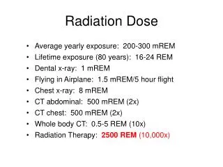
Head CT 1 - 2 mSv Chest CT 5 - 7 mSv Abd & Pelvis CT 8 - 11 mSv Average U.S. background radiation per year 3.6 mSv Typical chest X ray ~ 0.1 - 0.2 mSv Typical Effective Dose Values for CT Ca-Scoring 1.5 - 5.0 mSv Cardiac CTA 10 - 25 mSv Lecture 11: Cardiac CT - radiation doses, dose management and practical issues
Why doses are high in MDCT? • Shorter scan times and thinner slices requires higher tube current to maintain good image quality • For cardiac CT, excessive tissue overlap (low pitch) is often required to reduce motion artifacts • Translates to higher patient dose! Lecture 11: Cardiac CT - radiation doses, dose management and practical issues
Factors influencing MDCT radiation dose and image quality • Tube current (mA) • X ray on time • Pitch • mAs or effective mAs • X ray beam energy (kVp and filtration) • Slice thickness • Geometric and detector efficiency • Beam filters • Reconstruction algorithms, … • Patient size Lecture 11: Cardiac CT - radiation doses, dose management and practical issues
Dose (at the detector) vs. Noise 20 nGy per frame 150 nGy per frame 240 nGy per frame Lecture 11: Cardiac CT - radiation doses, dose management and practical issues
Effect of X ray beam energy 120 kVp 135 kVp Lecture 11: Cardiac CT - radiation doses, dose management and practical issues
Effect of Pitch on Dose and Image Quality P = 0.64 CTDI = 47.8 mGy 30% higher P = 0.83 CTDI = 37 mGy P = 1.48 CTDI = 20.6 mGy 45% lower Lecture 11: Cardiac CT - radiation doses, dose management and practical issues
CT Dosimetry CTDIw CTDI100 MSAD Effective Dose CTDIvol DLP Lecture 11: Cardiac CT - radiation doses, dose management and practical issues
Experimental Setup Radcal 1015C Electrometer, with CT ion chamber in body phantom Lecture 11: Cardiac CT - radiation doses, dose management and practical issues
Typical dose distribution in CT Body 32 cm Head16 cm 100 100 100 50 100 100 100 100 100 100 Dose uniform on surface and decreases towards center Lecture 11: Cardiac CT - radiation doses, dose management and practical issues
CTDIW(weighted average of center and periphery doses) CTDIw=(2/3) CTDIedge+(1/3) CTDIcenter CTDIvol (Pitch factor is considered) CTDIvol=(1/pitch)CTDIw Lecture 11: Cardiac CT - radiation doses, dose management and practical issues
Dose Length Product (DLP) • Indicates radiation dose of entire CT exam • Includes number of scans and scan width • DLP = CTDIvol (mGy) • scan length(cm) • Displayed on monitors Lecture 11: Cardiac CT - radiation doses, dose management and practical issues
Effective doses for Cardiac Imaging *Hunold P, et al., Radiology, 2003 Lecture 11: Cardiac CT - radiation doses, dose management and practical issues
Recent Advances: CT Dose Reductions Higher attenuation high mA Low attenuation low mA • X ray attenuation lower in AP and higher in lateral projection • However, CT doses are uniform on the surface and decrease radially towards center • Various dose reduction options are being considered Lecture 11: Cardiac CT - radiation doses, dose management and practical issues
Dose Reduction Options 200 mA 180 mA 150 mA 130 mA 150 mA 180 mA 210 mA 200 mA 170 mA • Dose reduction based on patient anatomy • Lower mA in AP, higher mA in lateral directions Methods • Patient attenuation measured during scout scan (AP & Lat) and alter mA for each gantry rotation (Smart mA1, Real AEC2) or “on-the-fly” (Care dose3) • Dose reduction of 20-40% is possible 1 GE, 2 Toshiba and 3 Siemens MDCT Lecture 11: Cardiac CT - radiation doses, dose management and practical issues
ECG-controlled tube current modulation • Tube current monitoring ECG signal is lowered in systole region and ramped up during diastole region • Mean radiation reduction of up to 45% has been reported* *Jakobs, et.al. Euro Radiol, 2002 Lecture 11: Cardiac CT - radiation doses, dose management and practical issues
Impact of Dose Modulation: Chest CT Radiation dose: Lateral: 16% increase, AP: 25% reduction *Mahesh, Kamel & Fishman, Evaluation of ‘CareDose’ on Siemens Sensation 64 MDCT scanner Lecture 11: Cardiac CT - radiation doses, dose management and practical issues
Lecture 11: Cardiac CT - radiation doses, dose management and practical issues
Dual Source CT* (DSCT) Tube A Tube B • Two X ray tubes positioned at right angle • Two detector arrays opposite to X ray tubes • Temporal resolutions less than 100 ms is possible by combining data from one-fourth of data acquired by two detectors *Siemens ‘Definition’ at Johns Hopkins, 2006 Lecture 11: Cardiac CT - radiation doses, dose management and practical issues
Conclusions • Radiation dose estimates for CT exams are best expressed as CTDIvol (mGy), DLP (mGy.cm) and Effective Dose E (mSv). • With increasing number of CT scans, there is concern about radiation burden to general public. Lecture 11: Cardiac CT - radiation doses, dose management and practical issues
Conclusions • Physicians referring or performing cardiac CT exams should understand radiation doses associated with various protocols and should be able to justify the appropriateness of CT exam • “Genie is out of the box, it is now left to the user how to tame it” Lecture 11: Cardiac CT - radiation doses, dose management and practical issues
Answer True or False • Typical patient dose values for cardiac CT angiography are in the range of 1-2.5 mSv. • Shorter scan times and thinner slices require higher tube current to maintain good image quality. • Patient dose increases if pitch factor increases (if all other parameters are maintained constant). Lecture 11: Cardiac CT - radiation doses, dose management and practical issues
Answer True or False • When patient radiation dose for CT is given as 500 mGy•cm, it is understood that skin dose is 500 mGy. • During CT cardiac angiography, tube current monitoring ECG signal is lowered in systole region and ramped up during diastole region. Lecture 11: Cardiac CT - radiation doses, dose management and practical issues