CT Radiation Dose Reduction
OPTIONAL LOGO HERE. CT Radiation Dose Reduction Pritesh Patel, M.D., Josh Weiss, D.O., Kevin Kirshenbaum, M.D., Mai Britt Campbell, M.D. Department of Radiology, Advocate Illinois Masonic Hospital, Chicago, IL. Why Minimizing Dose Is Important. ACR PQI Project. Learning Objectives.

CT Radiation Dose Reduction
E N D
Presentation Transcript
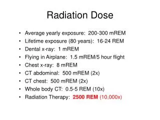
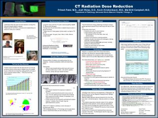
OPTIONALLOGO HERE CT Radiation Dose Reduction Pritesh Patel, M.D., Josh Weiss, D.O., Kevin Kirshenbaum, M.D., Mai Britt Campbell, M.D.Department of Radiology, Advocate Illinois Masonic Hospital, Chicago, IL Why Minimizing Dose Is Important ACR PQI Project Learning Objectives • Understand where the push for dose reduction is coming from. • Discuss biological effects of radiation. • Realize what the ACR PQI is, and how we are implementing it to reduce CT doses at our institution. • Long-term cancer risk from CT scans is deemed significant (BEIR VII, Brenner and Hall paper). • Average risk is 5% per Sievert (Sv) for radiation-induced cancer in general population. • Children are at 2-3 times greater risk than adults (as high as 15% per Sv). • For persons aged > 50 years, risk is 1/5th to 1/10th of that for younger adults. • The ACR (American College of Radiology) developed a practice quality improvement project (PQI) to help reduce the dose of Chest CTs. • Components of the Project: • Complete pre-survey of current practices • Conduct practice audit of 25 charts • We evaluated 10 CTs of the head, chest, and abdomen/pelvis. • Compare current practice to national guidelines • Complete education interventions (for radiologists, referring physicians, and technologists) • Create and implement action plan (see below), including deciding on clinical intervention. • Conduct six month follow up chart-review • Complete wrap up survey • Objective: to determine which of the clinical interventions described in the literature as reducing radiation exposure to patients is satisfactory for our practice in that it not only reduces radiation exposure but provides diagnostically acceptable images. • Our clinical interventions: (red = active, green = planned) • In plane bismuth breast and gonad shields • Reduce the tube voltage (kVp) • Reduce tube current or scan time (mAs) • Appropriate patient centering • Adjust CT imaging parameters based on patient height/weight. • Record patient doses incurred during diagnostic CT imaging studies. • Reduce scan range • Important additions to original ACR plan: • Looking at CTs of the head and abdomen/pelvis in addition to Chest CT • Addition of other clinical and technical interventions • Increase Pitch, Increase slice thickness • Testing/gathering data on all vendor CT Scans. • Pre and post-presentation test to gauge improvement. Fig 1: Various patients demonstrating deterministic radiation injury from unmonitored CT brain perfusion scans.2 Fig. 5 – Log used to acquire data. Initial Results • Data showed significant decreases in the CT Dose Index per volume (CTDIvol) and the Dose Length Product (DLP), both measures of overall dose to patient, while maintaining image quality. Our Action Plan Biological Effects of Radiation • Biological effects of radiation are broadly grouped into two categories – stochastic (probabilistic) effects and deterministic effects • Deterministic: • There is a dose threshold below which injury will not occur and above which injury is certain - skin injuries, epilation, cataracts, sterility, and acute radiation sickness The Push for Dose Reduction "If patient doses are higher than the expected level, but not high enough to produce obvious signs of radiation injury, the problem may go undetected and unreported, putting patients at increased risk for long-term radiation effects.“ – FDA (Food and Drug Administration1) • Educational Intervention Results: • Average post-lecture test scores were 87%. This was an approximately 20% improvement over pre-lecture scores. • As of March 2011, data has been acquired on many of the clinical interventions listed previously. In the following year, we plan on implementing additional interventions listed in the Action Plan, and evaluating subsequent effect on CT dose. Future Goals/Plans • Stochastic: • Risk of cancer induction from exposure to radiation is stochastic. • There is no dose threshold below which cancer does not occur. The risk of getting cancer increases with increasing dose, however the severity of illness (if it manifests) is not related to the dose received. • Certain cancers are more frequently associated with radiation exposure - leukemia, thyroid, breast, lung, and gastrointestinal tract cancers. • Effect of CT irradiation is primarily a concern because of stochastic-type effects. • With increasing advances in CT technology and new applications, however, deterministic effects are also becoming a concern. Fig 4 Summary of Methods • A Radiation Dose Task Force was established of Radiologists, the Medical Physicist, and the Diagnostic Imaging Director, Manager, and Clinical Specialist. • Baseline dosage data was collected in April 2010 for CTs of the head, chest, and abdomen/pelvis. Data was entered on a log (see Fig 5) • The residents presented an education session for the radiology department on dose reduction, and an exam was administered. • CT Chest protocol kilovoltage was reduced from120kv to 100kv. • Slice thickness on the CT Abdomen/Pelvis protocol was increased from 1.25mm to 2.5mm. • Another set of CT Chest and CT Abdomen/Pelvis scans were evaluated in November. The Radiologists rated the image quality on the lower dose scans. Fig 2: Bar graph indicating exponential growth in number of CT scans since 1993, approaching 70 million as of 2006. References/Acknowledgements Fig 3: Pie chart shows 46% increase in proportion of CT attributable dose from 1980 to 2006. Numbers are probably more pronounced today.