Download
1 / 50
610 likes | 1.27k Vues
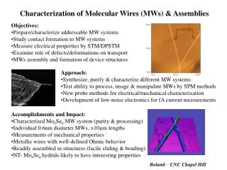
Molecular Characterization of Pasteurella multocida Isolates of Clinical significance. By RABIA DURRANI Department of Microbiology QUAID-I-AZAM UNIVERSITY, ISLAMABAD. CONTENTS. Introduction. Objectives of Study. Materials and Methods. Results. INTRODUCTION. Focus of the study

E N D
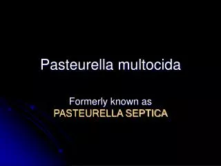
Molecular Characterization of Pasteurella multocida Isolates of Clinical significance By RABIA DURRANI Department of Microbiology QUAID-I-AZAM UNIVERSITY, ISLAMABAD.
CONTENTS Introduction Objectives of Study Materials and Methods Results
INTRODUCTION Focus of the study Disease and it’s causative agents
Livestock and Poultry sectors are among the major contributors in the economic growth of Pakistan Both sectors are facing serious problems; among them are some dangerous diseases like Hemorrhagic Septicemia (HS) which are posing a huge risk to the economic development in these sectors The focus of the research was to study the disease and identify the agents responsible for. Focus of the study
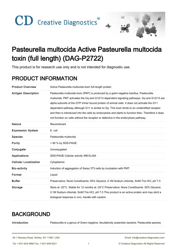
Disease and it’s causative agent(s) Pasteurella multocida is Gram negative, Non-spore forming, cocobacillus bacterium Commensals of upper respiratory tract of livestock and Poultry Causes severe Hemorrhagic septicemia (HS) HS occurs as catastrophic epizootics in Asia and Africa
Several cases of HS reported in Wild animals also. • Common serotypes are Asian B:2 and African E:2 • In wild animals B:2,5 strain is most common. • Vaccination is achieved by Whole cell preparations. • Common Vaccines are alum precipitated and Oil adjuvant. Disease and it’s causative agent(s) (Continued….)
Live Intranasal Vaccine B:3,4 serotype of deer origin found successful in South East Asia. (Muneer and Afzal, 1989). • Common cause of HS disease is Vaccination Failure and that is why death rate of animals is High in South East Asia. Disease and it’s causative agents (Continued….)
Objectives of Study Comparisons of isolates from clinically diseased animal (animal suffering from Hemorrhagic Septicemia) for their microbiological profiles to explore any gross difference. Study of molecular profiles to elucidate further proteomics. To collate and analyze the data obtained and to examine perception of the scientific workers that Pasteurella multocida is an opportunistic pathogen.
Sample collection & Identification of clinical isolates • Antibiogram analysis of Pasteurella multocida • Molecular Characterization of P. multocida • P. multocida species specific PCR • HS-associated type B serotype specific PCR • Protein Profile Analysis of P. multocida by SDS-PAGE • Whole Cell Profile of P. multocida • Envelop profile of P. multocida • Restriction enzyme analysis of P. multocida MATERIALS AND METHODS
Samples were collected from National Veterinary Labs Islamabad that were brought into the lab from different cities • Nature of the samples were lungs and spleen tissues of animals. • Gram staining of Organism • Subculturing and Purification • Biochemical characterization • Catalase test • Oxidase test • Growth in the absence of CO2 Sample Collection and Identification of clinical isolates
Urease production test • H2S Production Test • Nitrate reduction test • Motility test • Injecting mice with clinical isolates of clinical Isolate • API 20 NE Test (Biomeurix ) • Hyaluronidase Enzyme test for Pasteurella multocida (Smith et al., 1968) Identification of clinical isolates (Continued….)
Antibiogram analysis of Pasteurella multocida • Differentiation of clinical isolates by microbiological staining dyes: • Crystal violet • Malachite Green • Safranin • Congo Red
Molecular Characterization of Pasteurella multocida • Crude method of DNA extraction • Amplification of Extracted DNA of Pasteurella multocida by Polymerase Chain Reaction (PCR) • P. multocida species specific PCR assay
Primer sequences were: • KMT1SP6 5’ - GCTGTAAACGAACTCGTCGTCGCCAC3-3’ • KMT1T7 5’- ATCCGCTATTTACCCAGTGG-3’ P. multocida species specific PCR
PCR Master Mix Preparation • PCR master mix about 20µl was prepared as follows: • H2O 13µl • MgCl2 02µl • dNTPs 01µl • Primer 1 01µl • Primer 2 01µl • PCR Buffer 1.5µl • Taq Polymerase 0.5µl • Total Volume: 20µl • Then 5µl –extracted DNA was added, and makes the final volume upto 25µl.
The thermal cycling parameters were as follows: The initial denaturation at 94°C for 5minutes; • 30cycles of: • 94°C for 1minute • 53°C for 1minute (for primer annealing) • 72°C for 1minute (extension) • Final extension at 72°C for 9minutes.
Analysis of PCR product • About 8µl of PCR product were separated by electrophoresis on 2% High melting Agarose gel in 1X Tris-Boric acid running buffer (TBE) at 4V/cm for 1 hour .The gel was stained with 1% ethidium bromide and DNA fragments were viewed by Gel documentation system.
PCR assay for HS-associated type B serotype Pasteurella multocida • PCR analysis for HS –associated type B serotype P. multocida identification was performed. • Primer pair used: • KTSP61 • KTT72 • These primers specifically amplify a product of approximately 560 base pair (bp) in all HS causing serotype of P. multocida.
Primer sequences were: • KTT72 5’- AGGCTCGTTTGGATTATGAAG-3’ • KTSP61 5’ – ATCCGCTAACACACTCTC-3’ HS causing Type B gene specific PCR test
PCR master mix preparation PCR master mix about 20ul was prepared as follows: • H2O 13µl • MgCl2 02µl • dNTPs 01µl • Primer 1 01µl • Primer 2 01µl • Buffer 1.5µl • Taq Polymerase 0.5µl • Total Volume: 20µl • Then 5ul of extracted DNA was added, and make the final volume upto 25µl.
The thermal cycling parameters were as follows: • The initial denaturation at 94°C for 5minutes; • 30cycles of: • 94°C for 1minute • 53°C for 1 minute • 72°C for 1minute • Final extension at 72°C for 9minutes.
Protein Profile Analysis of P. multocida by SDS-PAGE • Harvesting of Culture • Washing of Cells • Sonication (Disruption) of Cells for Whole Cell Preparation • Preparation of Whole-cell for SDS-PAGE • Preparation of Envelope • Staining and Destaining of Gel • Determination of Molecular Weight of Proteins
For the extraction of proteins, thick growth of pure P. multocida were taken with the help of Pasteur pipette and dipped in the ependorff tube containing 200µl-distilled water. • Then they were vortexed with the help of (IKA USA mixer) thoroughly to dissolve completely. • After dissolving the samples were sonicated in (dr.hielscher, type UP 400S). Whole Cell Profile of Pasteurella multocida
Sonicated for: - • 30 second stroke • 30 second cooling • Amplitude 80 to 100 • Cycle 0.5 to 1 • After sonication: • centrifugation at 12,000 rpm • Time 5 min • The microcentrifuge was used to remove intact cells. The supernatant was taken as whole cell and boiled in boiling water along with 4X sample loading buffer for 5minutes at 100°C before loading on gel
High Energy Probe Sonicator (DR. HEILSCHER Type 2005, Germany)
Preparation of Envelope (Irfan et al.,2008) • Thick growth of P. multocida culture was: • Suspended in 2ml of distilled water in eppendorff tube • Centrifuged at 20,000rpm for 30minutes at 8˚C. • The pellet was resuspended in 20mM Tris-HCl buffer and again centrifuged at 20,000rpm for 30minutes. • The final pellet rich in envelop was resuspended in 20mM Tris-HCl buffer and stored at –20°C for further SDS-PAGE analysis. Envelop profile of P.multocida
Steps Involved • 10 X restriction endonuclease buffer • DNA • Distilled water • Restriction enzyme and then gently mix the contents • Centrifuge for few seconds in a microfuge and the incubate at 37°C for 1-4 hours • Heat the reaction at 65°C for 20 minutes • Run the digests on gel Restriction enzyme analysis of P.multocida
RESULTS Characterization of P.multocida Cultural and microscopic Biochemical Analytical Profile Index (API) test Antibiotic sensitivity Hylouronidase production Differentiation by microbiological staining dyes PCR SDS-PAGE
Cultural Characterization of P.multocida • On Brain Heart Infusion (BHI) agar and Nutrient agar: • Convex colonies • Entire edges • Mucoid and sticky nature • Approximate size of 2-3 mm diameter • On blood agar, all the pure culture showed: • Luxuriant growth • Translucent grayish or yellowish green colonies • No haemolysis • On MacConkey agar: • No growth Staining of P.multocida • The Gram staining shows: • Pleomorphic, gram negative, coccobacilli • 0.2-0.4 mm in size
Table 1. Common biochemical properties of Pasteurella multocida
Table 2. Analytical Profile Index (API) test results of Pasteurella multocida
Table 3. Number of clinical isolates sensitive to the antibiotics
Rapid plate screening test for Hyaluronidase production of P.multocida
Differentiation of Clinical strains of P.multocida by different staining dyes
Table 5. Differentiation of Clinical isolates by Microbiological staining dyes
Amplification of Extracted DNA of Pasteurella multocida by PCR • All samples when run on Agarose Gel Electrophoresis showed: • Bands of base pairs 560 and 750 • The DNA ladder started from 100 base pairs.
Organism was genetically conserved and there is no demostratable diversity with in the number of samples tested. • Isolates from wild life be included in such studies. • This organisms maintains harmlessly inhealthy animals and causes disease as an opportunistic pathogen in extreme stress to animals. • By further studies we can devise better methods for control against disease caused by this organism. Conclusions & Future Prospect