Download
1 / 33
340 likes | 514 Vues
B – CELL ACTIVATION Where and how do all these things take place?. B cells in blood. T cell area. B cell area. Efferen s lymph. B-cell recycling in the absence of antig e n ( lymph node ). B cells proliferate rapidly. B cells leave blood & enter lymph node via

E N D
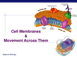
B – CELL ACTIVATION Where and how do all these things take place?
B cells in blood T cell area B cell area Efferens lymph B-cell recycling in the absence of antigen (lymph node)
B cells proliferate rapidly B cells leave blood & enter lymph node via high endothelial venules Antigen enters node in afferent lymphatic Y Y Y Y Y Y Y Y Y Y Y Y Y Y Germinal centre releases B cells that differentiate into plasma cells Y Y Y Y GERMINAL CENTRE Transient structure of Intense proliferation Recirculating B cells are trapped by foreign antigens in lymphoid organs
Antigen is bound on the surface of follicular dendritic cells (FDC) FDC FDC bind immune complexes (Ag-Ab ) Ag detectable for 12 months following immunization A single cell binds various antigens FDC depends on the presence of TNF-α, LTα, LTβ B cells recognize Ag on the surface of FDC On the surface of FDC immune complexes form the so-called iccosomesthat can be released and taken up later by the surrounding germinal center B cells
The structure of the germinal centre Somatic hypermutation LZ FDC DZ Somatic hypermutation LZ: light zone DZ: dark zone FDC: follicular dendritic cell
B B B B B B B B B B IMMUNOLOGICAL MEMORY – B CELLS • Germinal Centre reaction • proliferation • somatic hypermutation • affinity maturation • Memory B cells • previously activated • passed affinity maturation • present in the circulation • rapidly proliferate and differentiate to plasma cell upon re-activation, or enter the GC reaction again • Plasma cells • provides serological memory: • pre-existing neutralizing Abs to pathogens or toxins B B FDC B FDC B T B plasma cell T B
Selection Repeated cycle Somatic hypermutation
T CELL DEPENDENT B CELL ACTIVATION IN LYMPHOID ORGANS BLIMP expresszió B-lymphocyte induced maturation protein IgM IgG IgA IgE
Regulation of memory vs plasma cell differentiation B cell T cell - activated Bcl-6 szuppresszor BLIMP gátló CD40 signaling
T B CD40L CD40 Follicular dendritic cell (FDC) B CD21 FcR Ag Memory B cell FcR B NO Ag apoptosis DEVELOPMENT OF B CELL MEMORY IN THE FOLLICLE
CELL INTERACTIONS IN PERIPHERAL LYMPHOID TISSUES T cell area – PALS paracortex Germinal center DC – T cellcontact DC Proliferating B cells centroblasts T B B – T cell contact Somatic hypermutation Additional gene rearrangement Isotype switch Plasma cell differentiation Antibody production Marginal zone Memory B cells Arteriole
Tight junction B cell VLA-4 VCAM-1 Inhibition of apoptosis ICAM-1 LFA-1 BCR C3d CD21 Follicular dendritic cells SELECTION OF HIGH AFFINITY B CELLS UPON INTERACTION WITH FOLLICULAR DENDRITIC CELLS
FDC apoptosis Fas B cell FasL differentiation GC T cell CD40 CD40L INTERACTION OF ANTIGEN-SPECIFIC T AND B CELLS B and T cells recognize the same antigen
How antigen-specific Ab production is maintained? MODEL 2. MODEL 1. memory B cell plasma cell Bystander help: Cross-reactive antigens TLR ligands Cytokines... Memory B cells continuously differentiate into plasma cells Long-lived plasma cells in the bone marrow MODEL 3. • Repeated activation with the antigen drives B cell activation and plasma cell differentiation • role of follicular dendritic cells in antigen storage (months-years?) • Polio: reinfections with Sabin vaccine strain • subclinical infections (Diphteria in 10% of the population) • hidden antigens (Measles genes persist in neurons – can induce Subacute Sclerosing Panencephalitis)
CD40 CD40L MHC TCR DC T cell CD40L TCR CD28 B7 CD28 CD40 MHC B7 B cell CELL INTERACTIONS IN THE PARACORTEX Antigen recognition by B and T lymphocytes
Ligand Ligand SIGNAL SIGNAL RECEPTOR MEDIATED CELL ACTIVATION Cross - linking Conformational change
CROSS – LINKING OF THE RECEPTOR INITIATES A SIGNALING CASCADE ligand kinase activation phosphorylation recruitmentof adaptors Activation of transcription factors Gene transcription SIGNAL
a a antigen binding V V V V L L mIg molecule Kinases H H Signal transduction b SHP-1 a Syk Phosphatases Btk PLC Vav Lyn HS1 Adaptors + substrates SLP-65/BLNK THE IgM B-CELL RECEPTOR Ig-a/Ig-b heterodimer
Ig domain + CHO a b ITAM ITAM Y Y Y Y SIGNALING UNITS OF THE B-CELL RECEPTOR Ig-a/CD79a Ig-b/CD79b ITAM: YxxLx7YxxI ITAM: Immunoreceptor Tyrosine-based Activation Motif
Ag 2. Src-family kinase activation and ITAM phosphorylation 3. Syk recruitment and activation 4. SLP phosphorylation+Ca release Syk Syk Lyn Lyn SLP P P P P P P P Calcium release = ITAM RECENT MODEL OF B-CELL RECEPTOR MEDIATED SIGNALING Subsequent activation of 2 kinases 1. Cross-linking
THE CO-STIMULATORY ROLE OF CR2 (CD21) COMPLEMENT RECEPTOR IN B – LYMPHOCYTES C3d ANTIGEN Antigenic determinant CD21/CR2 CD19 TAPA=CD81 Y Y B-CELL Enhanced B-cell activation
Mannose Tissue cells Bacterium Antigen B Cell THE NEURAMIC ACID RECEPTOR CD22 INHIBITS ACTIVATION THROUGH THE A B-CELL RECEPTOR Neuraminic acid CD22 Inhibited B cell activation
KINETICS OF LYMPHOCYTE ACTIVATION Nyugvó limfocita G0 Resting lymphocyte G0 Ko-receptor Adhesion molecule Cytokines SIGNAL2. Effector cellMemory cell Transport Membrane change RNA and protein synthesis sejtosztódás DNA synthesis Lymphoblast PTK activation RNA synthesis Free Ca++ Protein synthesis Protein phosphorylation DNA synthesis Resting lymphocyte G0 0 10sec 1min 5min 1hr 6 hrs 12 hrs 24 hrs ANTIGEN SIGNAL1.
Neutralization – binding of the antibody inhibits the adhesion of the pathogen, its entry or multipolication Opsonization – binding of antibodies induces complement activation and promotes binding to immun cells through complement (CR1) and immunoglobulin binding (FcR) receptors Antibody isotypes differ in their complement activating and FcR binding capabilities ANTIBODY – METIDATED EFFECTOR FUNCTIONS
OPSONIZATION Binding of antibody increases phagocytosis FcR COMPLEMENT ACTIVATION Opsonization by C3b PLASMA CELL Complement C3b FcR FcR CR1 EFFECTOR FUNCTIONS OF ANTIBODIES INHIBITION Binding of bacteria to epithelial cells Binding of viruses to receptor Binding of bacterial toxins to target cells NEUTRALIZATION Small proportion of antibodies PHAGOCYTES ENGULFMENT, DEGRADATION
Models of Human Rhinovirus 14 neutralised by monoclonal antibodies 30nm Human Rhinovirus 14 - a common cold virus 30 strongly neutralising McAb 60 strongly neutralising McAb Fab regions 60 weakly neutralising McAb Fab regions
Electron micrographs of Antibodies and complement opsonising Epstein Barr Virus (EBV) Negatively stained EBV EBV coated with antibodies and activated complement components EBV coated with a corona of anti-EBV antibodies
T – CELLS PROMOTE B – CELL DIFFERENTIATION ANTIGEN CYTOKINES PLASMA CELL B -CELL ISOTYPE SWITCH AND AFFINITY MATURATION OCCURS IN COLLABORATION WITH T – CELLS ONLY HOW T – CELLS RECOGNIZE ANTIGENS?