Download
1 / 44
450 likes | 790 Vues
Responses to Resistance training (Anaerobic Training). Key Term . anaerobic training: High-intensity, intermittent bouts of exercise such as weight training; plyo -metric drills; and speed, agility, and interval training. Table 5.2. ( continued ). Table 5.2 (continued). ( continued ).

E N D
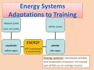
Key Term • anaerobic training: High-intensity, intermittent bouts of exercise such as weight training; plyo-metric drills; and speed, agility, and interval training.
Table 5.2 (continued)
Table 5.2 (continued) (continued)
Neural Adaptations • Anaerobic training may elicit adaptations along the neuromuscular chain, beginning in the higher brain centers and continuing down to the level of individual muscle fibers.
Neural Adaptations • Central Adaptations • Motor cortex activity increases when the level of force developed increases and when new exercises or movements are being learned. • Many neural changes with anaerobic training take place along the descending corticospinal tracts. • Adaptations of Motor Units • Maximal strength and power increases of agonist muscles result from an increase in recruitment, rate of firing, synchronization of firing, or a combination of these factors.
Key Point • With heavy resistance training, all muscle fibers get larger because they are all recruited in consecutive order by their size to produce high levels of force. In advanced lifters, the central nervous system might adapt by allowing these athletes to recruit some motor units not in consecutive order, recruiting larger ones first to help with greater production of power or speed in a movement.
Size Principle • Figure 5.2 (next slide) • The slide shows a graphic representation of the size principle, according to which motor units that contain Type I (slow-twitch) and Type II (fast-twitch) fibers are organized based on some “size” factor. • Low-threshold motor units are recruited first and have lower force capabilities than higher-threshold motor units. • Typically, to get to the high-threshold motor units, the body must first recruit the lower-threshold motor units. • Exceptions exist, especially with respect to explosive, ballistic contractions that can selectively recruit high-threshold units to rapidly achieve more force and power.
Neural Adaptations • Neuromuscular Junction • Possible changes with anaerobic training include • increased area of the neuromuscular junction (NMJ); • more dispersed, irregularly shaped synapses and a greater total length of nerve terminal branching; and • increased end-plate perimeter length and area, as well as greater dispersion of acetylcholine receptors within the end-plate region. • Neuromuscular Reflex Potentiation • Anaerobic training may enhance the reflex response, thereby enhancing the magnitude and rate of force development.
Neural Adaptations • Anaerobic Training and Electromyography (EMG) Studies • An increase in EMG indicates greater neural activation. • Studies have shown strength and power increases of up to 73%. • Advancement in training contributes to further gains in strength and power. • Dramatic increases in neural adaptations take place early in the training program. • Additional findings include the following: • Cross-education • Bilateral deficit in untrained individuals • Changes in muscle activity of the antagonists during agonist movements
Neural Adaptations w Synchronization and recruitment of additional motor units • Decreased neural inhibition; e.g., decreased GTO effects w Decreased co-activation of antagonist muscles w Increased rate coding (increased firing frequency of active motor units) Muscle Hypertrophy w Fiber hypertrophy w Fiber hyperplasia (??? – probably not) Mechanisms of Gains in Muscle Strength
Results of Resistance Training on Muscle Strength in Males w Alterations of neural control of trained muscle. w Increased muscle size (hypertrophy).
Muscle Fiber Hypertrophy w The numbers of myofibrils and thick and thin filaments increase, so there are more cross-bridges in the cross-section of muscle, and hence, greater strength. wProtein turnover is continuous in the muscle; during hypertrophy, muscle protein synthesis increases more than proteindegradation during the post-exercise period. wTestosterone plays a role in promoting muscle growth. w Training at higher intensities, i.e., performing lower reps with higher loads, appears to cause greater fiber hypertrophy than training at lower intensities.
Key Point • The process of hypertrophy involves both an increase in the synthesis of the contractile proteins actin and myosin within the myofibril and an increase in the number of myofibrils within a muscle fiber. The new myofilaments are added to the external layers of the myofibril, resulting in an increase in its diameter.
Muscular Adaptations • Fiber Size Changes • Resistance training results in increases in both Type I and Type II muscle fiber area. • Type II fibers have greater increases in size than Type I fibers. • Fiber Type Transitions • There is a continuum of fiber types: I, Ic, IIc, IIac, IIa, IIax, IIx.
Muscle Fiber Transitions • Figure 5.3 (next slide) • Muscle fiber transitions occur during training. • This means that a shift of the type of myosin adenosine triphosphatase (ATPase) and heavy chains takes place during training. • Transformations from IIx to IIax to IIa can be seen, and then small percentages change to IIac and IIc. • Exercise activities that recruit motor units with Type IIx muscle fibers initiate a shift toward IIa fibers.
Muscular Adaptations • Structural and Architectural Changes • Resistance training increases myofibrillar volume, cytoplasmic density, sarcoplasmic reticulum and T-tubule density, and sodium-potassium ATPase activity. • Sprint training enhances calcium release. • Resistance training increases angle of pennation. • Other Muscular Adaptations • Reduced mitochondrial density • Decreased capillary density • Increased buffering capacity (acid-base balance) • Changes in muscle substrate content and enzyme activity
Muscle Fiber Hyperplasia w Muscle fibers may split with intense weight training. w Each half may then increase in size. w Hyperplasia has been shown to occur in some experimental animal models; it has not been clearly demonstrated in human subjects. Quail and chicken: hanging a weight on the wing – hypertrophy of the latissimus muscles Cat: pulling a lever to obtain food (not as clear) – hypertrophy of the wrist flexor muscles
Perry and Rudnicki (2000) Frontiers in Bioscience 5:D750-67. 4 days after damage 2 weeks after damage 4 weeks after damage with irradiation Repair through activation of satellite cells Myology (Sanes, McGraw-Hill, 1994)
Myonuclei and satellite cells Hawke, Exerc Sport Sci Rev 33: 63, 2005
2 weeks post * p < 0.05 * * A single prior exposure to a protocol of lengthening contractions reduced the force deficit and damaged fibers 60% non-trained 50 40 30 20 10 0 Force Deficit (% control) Injured Fibers (% total) Koh & Brooks (2001) Am J Physiol 281:R155-R161.
60 60 50 50 40 40 30 30 20 20 10 10 0 0 Degeneration-regeneration not necessary to provide muscles protection from contraction-induced injury non-trained trained passive trained isometric *different from non- -trained (p<0.05) * * Force deficit (% control) Injured fibers (% total) * Koh & Brooks (2001) Am J Physiol 281:R155-R161. Force deficit Injured fibers • Despite the increase in susceptibility to injury with aging, • and the decreased ability to recover, muscles in old • animals can be conditioned for protection from injury. • Maintenance of conditioned fibers, particularly in muscles • of elderly people, may prevent inadvertent damage during • contractions.
Bone Modeling • Figure 5.4 (next slide) • (a) Application of a longitudinal weight-bearing force causes the bone to bend (as depicted by the dotted line), creating a stimulus for new bone formation at the regions experiencing the greatest deformation. • (b) Osteoblasts lay down additional collagen fibers. • (c) Previously dormant osteoblasts migrate to the area experiencing the strain. • (d) The collagen fibers become mineralized, and the bone diameter effectively increases.
Connective Tissue Adaptations • General Bone Physiology • Trabecular bone responds more rapidly to stimuli than does cortical bone. • Minimal essential strain (MES) is the threshold stimulus that initiates new bone formation. • The MES is approximately 1/10 of the force required to fracture bone.
Key Point • Forces that reach or exceed a threshold stimulus initiate new bone formation in the area experiencing the mechanical strain.
Connective Tissue Adaptations • Anaerobic Training and Bone Growth • Muscle strength and hypertrophy gains increasethe force exerted on the bones, which may result ina corresponding increase in bone mineral density (BMD) or the quantity of mineral deposited in agiven area of bone.
Connective Tissue Adaptations • Principles of Training to Increase Bone Strength • Magnitude of the load (intensity) • Rate (speed) of loading • Direction of the forces • Volume of loading (number of repetitions)
Connective Tissue Adaptations • How Can Athletes Stimulate Bone Formation? • Use exercises that directly load particular regions of the skeleton. • Use structural exercises to direct force vectors through the spine and hip and allow the use of greater absolute loads in training. • Overload the musculoskeletal system, and progressively increase the load as the tissues become accustomed to the stimulus. • Vary exercise selection to change the distribution of the force vectors to continually present a unique stimulus.
Key Point • Programs designed to stimulate new bone formation should incorporate the concepts of specificity of loading, proper exercise selection, progressive overload, and vari-ation. The exercises selected should be structural and weight bearing.
Connective Tissue Adaptations • Adaptations of Tendons, Ligaments, and Fascia to Anaerobic Training • The primary stimulus for growth of tendons, ligaments, and fascia is the mechanical forces created during exercise. • The degree of tissue adaptation is proportional to the intensity of exercise. • Consistent anaerobic exercise that exceeds the threshold of strain stimulates connective tissue changes.
Formation of a Collagen Fiber • Figure 5.5 (next slide) • The primary structural component of all connective tissue is the collagen fiber (Type I for bone, tendon, and ligaments and Type II for cartilage).
Connective Tissue Adaptations • Adaptations of Tendons, Ligaments, and Fascia to Anaerobic Training • Sites where connective tissues can increase strength and load-bearing capacity are • at the junctions between the tendon (and ligament) and bone surface, • within the body of the tendon or ligament, and • in the network of fascia within skeletal muscle.
Connective Tissue Adaptations • Adaptations of Tendons, Ligaments, and Fascia to Anaerobic Training • Specific tendinous changes that contribute to size and strength increases include • an increase in collagen fibril diameter, • a greater number of covalent cross-links within the hypertrophied fiber, • an increase in the number of collagen fibrils, and • an increase in the packing density of collagen fibrils.
Connective Tissue Adaptations • How Can Athletes Stimulate Connective Tissue Adaptations? • Tendons, Ligaments, Fascia • Exercise of low to moderate intensity does not markedly change the collagen content of connective tissue. • High-intensity loading results in a net growth of the involved connective tissues.
Connective Tissue Adaptations • Cartilage Adaptations to Anaerobic Training • The main functions of cartilage are to • provide a smooth joint articulating surface, • act as a shock absorber for forces directed through the joint, and • aid in the attachment of connective tissue to the skeleton.
Connective Tissue Adaptations • Cartilage Adaptations to Anaerobic Training • Cartilage lacks its own blood supply and must depend on diffusion of oxygen and nutrients from synovial fluid. • Therefore, joint mobility is linked with joint health. • Movement about a joint creates changes in pressure in the joint capsule that drive nutrients from the synovial fluid toward the articular cartilage of the joint.
Connective Tissue Adaptations • How Can Athletes Stimulate Connective Tissue Adaptations? • Cartilage • Weight-bearing forces and complete movement throughout the range of motion seem to be essential to maintaining tissue viability. • Moderate aerobic exercise seems adequate for increasing cartilage thickness. • Strenuous exercise does not appear to cause degenerative joint disease.