Download
1 / 37
410 likes | 717 Vues
بسم الله الرحمن الرحيم. Functional Anatomy of Heart. Dr.Mohammed Sharique Ahmed Quadri Assistant Professor Physiology Almaarefa College. FUNCTIONAL ANATOMY OF HEART. Heart beat starts in the fourth week after conception during intra-uterine life, when embryo is only few millimeters.

E N D
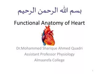
بسم الله الرحمن الرحيم Functional Anatomy of Heart Dr.Mohammed Sharique Ahmed Quadri Assistant Professor Physiology Almaarefa College
FUNCTIONAL ANATOMY OF HEART Heart beat starts in the fourth week after conception during intra-uterine life, when embryo is only few millimeters. Throughout life, heart keeps on beating and it contracts about 3 billion times during an average life span.
Circulatory System • Three basic components • Heart • Serves as pump that establishes the pressure gradient needed for blood to flow to tissues • Blood vessels • Passageways through which blood is distributed from heart to all parts of body and back to heart • Blood • Transport medium within which materials being transported are dissolved or suspended
CIRCULATORY SYSTEM • We have two types of circulation: i). Pulmonary Circulation ii). Systemic Circulation • Pulmonary Circulation Blood is carried from the right ventricle of the heart to lungs and back to left atrium of the heart. • Systemic Circulation Blood is carried from left ventricle to the body and back to the right atrium.
FUNCTIONAL ANATOMY OF HEART Heart is muscular organ. It is involuntary, present in the middle of the thoracic cavity, about the size of fist [14cm long, 9cm wide]. Sternum lies anteriorly and vertebral column [backbone] lies posteriorly and lungs laterally. Heart has base and apex. - Base is at the top, behind the 2nd intercostal space. - Apex is lower down in the 5th left intercostal space. Slightly below nipple towards mid line
FUNCTIONAL ANATOMY OF HEART • Applied • As the heart lies between the sternum and vertebral column, it is possible to compress the sternum and drive blood out of the heart when heart is not pumping effectively. • This external compression of heart is done in CPR [Cardio-Pulmonary Resuscitation], which is life saving, till proper therapy can be given.
Circulatory System • Heart IS A Dual pump • Right and left sides of heart function as two separate pumps • Divided into right and left halves and has four chambers • Atria • Upper chambers • Receive blood returning to heart and transfer it to lower chambers • Ventricles • Lower chambers which pump blood from heart
HEART AS A PUMP Right and left atrium are separated from each other by interatrial septum. Right and Left ventricle are separated by interventricular septum. [Septum is muscular wall which does not allow the blood to mix between two sides.]
HEART AS A PUMP In between atria and ventricle, there is fibrous ring, in which valves are embedded. Right atrium gets the blood from superior venaceva [SVC] and inferior venaceva [IVC].It is deoxygenated blood. Blood from right atrium goes to right ventricle through right AV valve or Tricuspid valve.
HEART AS A PUMP • From right ventricle, blood goes to pulmonary artery through pulmonary valve, to the lungs. • In the lungs, blood gets oxygenated and is returned to left atrium by 4 pulmonary veins. • IMPORTANT • Remember Pulmonary Artery is the only artery which carries deoxygenated blood and Pulmonary veins are only veins in the body which carry oxygenated blood.
HEART AS A PUMP From left atrium, blood goes to left ventricle through left AV valve or mitral valve [bicuspid valve]. From left ventricle, blood goes to aorta through aortic valve to the body.
LEFT PUMP COMPARISON OF RIGHT & LEFT PUMPS RIGHT PUMP • Both sides pump equal amount of blood. • Right side has deoxygenated blood [goes to lungs and gets O2 in the lungs] . • Pulmonary Circulation is low pressure circulation. • Pulmonary Circulation is low resistance circulation. • Right ventricle wall is thin [2-3mm]. • Both sides pump equal amount of blood. • Left side has oxygenated blood. • Systemic Circulation is high pressure circulation. • Systemic Circulation is high resistance circulation. • Left ventricle wall is thick [8-10mm].
HEART VALVES There are four valves. They are one way valve. Valves open and close passively because of pressure difference. Function of the valve is to prevent back flow of the blood.
HEART VALVES Four Valves are: Two AV [Atrioventricular] Valves i). Right AV Valve – Tricuspid Valve ii). Left AV Valve – Mitral or Bicuspid Valve Semi lunar Valves iii). Aortic Valve iv). Pulmonary Valve
AV VALVES AV Valves [Tricuspid & Mitral] are attached to the papillary muscle and chordae tendineae. Chordae tendineae are tendon like tissue and their function is prevent the eversion or bulging of valves into atria. Chordae tendineae are attached to papillary muscle which protrude from inner surface of ventricular valve. Important -- Papillary muscle and chordae tendineae are attached to AV valves only.
AV VALVES When ventricle contract, papillary muscle also contract and pull down the chordae tendineae, which keeps the valve tightly closed. There are three papillary muscles attached to the right AV valve [tricuspid valve]. There are two papillary muscle attach to the left AV valve [bicuspid or mitral valve].
SEMI-LUNAR VALVES • Aortic Valve and Pulmonary Valve • Aortic Valve is present at the beginning of aorta and has three cusps. • Pulmonary Valve is present at the beginning of pulmonary artery and has three cusps. • Aortic and Pulmonary Valve open when pressure increases in left and right ventricle during ventricular contraction. • They close when ventricular pressure decreases than aortic and pulmonary artery pressure. • They prevent back flow of the blood.
FIBROUS SKELETON OF THE HEART In between atria and ventricles, there is fibrous ring [it is dense connective tissue]. It provides base for attachment of four heart valves. Atrial Muscle is attached to upper part and ventricular muscle is attached to the bottom of the ring. Fibrous ring is non-conductive, therefore, special conductive tissue is required to conduct impulse from atria to ventricle.
LAYERS OF THE HEART Heart has 3 Layers: 1. Endothelium – inner lining of the heart 2. Myocardium – cardiac muscle tissue 3. Pericardium – external layer Pericardium has two layers inner visceral [called epicardium] and outer parietal layer. There is pericardial fluid about 5 – 30 ml present between two layers. It prevents friction between the layers as they move over each other with every beat of the heart.
APPLIED - PERICARDITIS Pericarditis is the inflammation of pericardium. It results in painful friction rub between the two layers of pericardium. It can be caused by viral or bacterial infection.
BLOOD SUPPLY TO THE HEART Heart is supplied by coronary arteries [branches of aorta]. Cardiac muscle fibers have rich blood supply, about 1 capillary for each myocardial fiber. Cardiac muscle has abundance of energy generating mitochondria.
AUTONOMIC NERVE SUPPLY Heart is supplied by sympathetic and parasympathetic nerves. Sympathetic Stimulation causes increased force of contraction [positive INOTROPIC effect] and increase heart rate [positive CHRONOTROPIC effect]. Parasympathetic Stimulation decreased force of contraction [negative INOTROPIC effect] and decrease in heart rate [negative CHRONOTROPIC effect].
CARDIAC MUSCLE MICROSCOPIC STRUCTURE Cardiac Muscle fibers are connected by membrane called ‘INTERCALATED DISC’. Intercalated Disc have gap junction. Gap Junction allow relatively free diffusion of ions, therefore, action potential travels from one cell to another easily. Therefore, cardiac muscle works as SYNCYTIUM [one unit], therefore, heart can be depolarized all at one time, it obeys all or none law.
CARDIAC MUSCLE MICROSCOPIC STRUCTURE We have Atrial Syncytium and Ventricular Syncytium. They contract as separate units as they are separated by non-conducting fibrous ring. The impulse travels from the atria to the ventricle by specialized conductive tissue.
References • Human physiology by Lauralee Sherwood, seventh edition • Text book physiology by Guyton &Hall,11th edition • Text book of physiology by Linda .s contanzo,third edition


![CARDIO-VASCULAR SYSTEM [CVS] FUNCTIONAL ANATOMY OF HEART](https://cdn1.slideserve.com/1739818/cardio-vascular-system-cvs-functional-anatomy-of-heart-dt.jpg)