Download

1 / 34
340 likes | 468 Vues
Explore the intricate details of heart anatomy including its size, location, coverings, layers, major vessels, chambers, valves, and circulation pathways. Understand the physiological significance of each component for optimal heart function.

E N D
Anatomy of the Heart
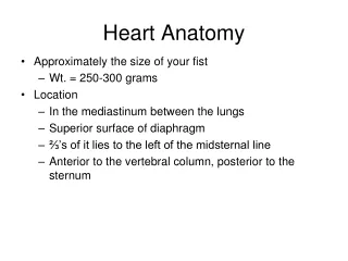
Heart Anatomy • Approximately the size of your fist • Location • Superior surface of diaphragm • Left of the midline • Anterior to the vertebral column, posterior to the sternum
Heart Anatomy Figure 18.1
Coverings of the Heart: Anatomy • Pericardium – a double-walled sac around the heart composed of: • A superficial fibrous pericardium • A deep two-layer serous pericardium • The parietal layer lines the internal surface of the fibrous pericardium • The visceral layer or epicardium lines the surface of the heart • They are separated by the fluid-filled pericardial cavity
Coverings of the Heart: Physiology • The pericardium: • Protects and anchors the heart • Prevents overfilling of the heart with blood • Allows for the heart to work in a relatively friction-free environment
Pericardial Layers of the Heart Figure 18.2
Heart Wall • Epicardium – visceral layer of the serous pericardium • Myocardium – cardiac muscle layer forming the bulk of the heart • Fibrous skeleton of the heart – crisscrossing, interlacing layer of connective tissue • Endocardium – endothelial layer of the inner myocardial surface
External Heart: Major Vessels of the Heart (Anterior View) • Vessels returning blood to the heart include: • Superior and inferior venae cavae • Right and left pulmonary veins • Vessels conveying blood away from the heart include: • Pulmonary trunk, which splits into right and left pulmonary arteries • Ascending aorta (three branches) – brachiocephalic, left common carotid, and subclavian arteries
External Heart: Vessels that Supply/Drain the Heart (Anterior View) • Arteries – right and left coronary (in atrioventricular groove), marginal, circumflex, and anterior interventricular arteries • Veins – small cardiac, anterior cardiac, and great cardiac veins
External Heart: Anterior View Figure 18.4b
External Heart: Major Vessels of the Heart (Posterior View) • Vessels returning blood to the heart include: • Right and left pulmonary veins • Superior and inferior venae cavae • Vessels conveying blood away from the heart include: • Aorta • Right and left pulmonary arteries
External Heart: Vessels that Supply/Drain the Heart (Posterior View) • Arteries – right coronary artery (in atrioventricular groove) and the posterior interventricular artery (in interventricular groove) • Veins – great cardiac vein, posterior vein to left ventricle, coronary sinus, and middle cardiac vein
External Heart: Posterior View Figure 18.4d
Gross Anatomy of Heart: Frontal Section Figure 18.4e
Atria of the Heart • Atria are the receiving chambers of the heart • Each atrium has a protruding auricle • Pectinate muscles mark atrial walls • Blood enters right atria from superior and inferior venae cavae and coronary sinus • Blood enters left atria from pulmonary veins
Ventricles of the Heart • Ventricles are the discharging chambers of the heart • Papillary muscles and trabeculae carneae muscles mark ventricular walls • Right ventricle pumps blood into the pulmonary trunk • Left ventricle pumps blood into the aorta
Pathway of Blood Through the Heart and Lungs • Right atrium tricuspid valve right ventricle • Right ventricle pulmonary semilunar valve pulmonary arteries lungs • Lungs pulmonary veins left atrium • Left atrium bicuspid valve left ventricle • Left ventricle aortic semilunar valve aorta • Aorta systemic circulation
Pathway of Blood Through the Heart and Lungs Figure 18.5
Coronary Circulation • Coronary circulation is the functional blood supply to the heart muscle itself • Collateral routes ensure blood delivery to heart even if major vessels are occluded
Coronary Circulation: Arterial Supply Figure 18.7a
Coronary Circulation: Venous Supply Figure 18.7b
Heart Valves • Heart valves ensure unidirectional blood flow through the heart • Atrioventricular (AV) valves lie between the atria and the ventricles • AV valves prevent backflow into the atria when ventricles contract • Chordae tendineae anchor AV valves to papillary muscles
Heart Valves • Aortic semilunar valve lies between the left ventricle and the aorta • Pulmonary semilunar valve lies between the right ventricle and pulmonary trunk • Semilunar valves prevent backflow of blood into the ventricles
Heart Valves Figure 18.8a, b
Heart Valves Figure 18.8c, d
Atrioventricular Valve Function Figure 18.9
Semilunar Valve Function Figure 18.10
Microscopic Anatomy of Heart Muscle • Cardiac muscle is striated, short, fat, branched, and interconnected • The connective tissue endomysium acts as both tendon and insertion • Intercalated discs anchor cardiac cells together and allow free passage of ions • Heart muscle behaves as a functional syncytium PLAY Cardiovascular System: Anatomy Review: The Heart
Microscopic Anatomy of Heart Muscle Figure 18.11