Download

1 / 13

E N D
Anatomy of the Heart BIOLOGY FORM 5
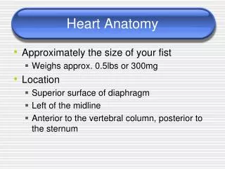
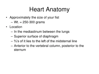
The heart is the organ that supplies blood and oxygen to all parts of the body. It is about the size of a clenched fist, weighs about 10.5 ounces and is shaped like a cone. The heart is located in the chest cavity just posterior to the breastbone, between the lungs and superior to the diaphragm. The heart is surrounded by a fluid filled sac called the pericardium. Blood is pumped away from the heart through arteries and returns to the heart through veins. The major artery of the body is the aorta and the major veins of the body are the vena cava.
Chambers • The heart is divided by a partition or septum into two halves. The halves are in turn divided into chambers. The upper two chambers of the heart are called atria and the lower two chambers are called ventricles. Valves allow blood to flow in one direction between the chambers of the heart.
What are atria? • The heart is divided into four chambers that are connected by valves. The upper two chambers of the heart are called the left atrium and right atrium. Function: • Right Atrium: Receives blood returning to the heart from the superior and inferior vena cava. • Left Atrium: Receives blood returning to the heart from the pulmonary veins
What are ventricles? • The heart is divided into four chambers that are connected by valves. The lower two chambers of the heart are called the left ventricle and the right ventricle. Function: • Right Ventricle: Receives blood from the right atrium and pumps it to the pulmonary artery. • Left Ventricle: Receives blood from the left atrium and pumps it to the aorta
What are heart valves? • Valves are flap-like structures that allow blood to flow in one direction. The heart has two kinds of valves, atrioventricular and semilunar valves. AtrioventricularValves • The atrioventricular valves are thin structures that are composed of endocardium and connective tissue. They are located between the atria and the ventricles. What is the mitral valve? • Valves are flap-like structures that allow blood to flow in one direction. The mitral valve is located between the left atrium and the left ventricle. Function: • Prevents the back flow of blood as it is pumped from the left atrium to the left ventricle.
SemilunarValves • The semilunar valves are flaps of endocardium and connective tissue reinforced by fibers which prevent the valves from turning inside out. They are shaped like a half moon, hence the name semilunar (semi-, -lunar).The semilunar valves are located between the aorta and the left ventricle and between the pulmonary artery and the right ventricle.
Heart Sounds • The audible sounds that can be heard from the heart are made by the closing of the heart valves. These sounds are referred to as the "lub-dupp" sounds. The "lub" sound is made by the contraction of the ventricles and the closing of the atrioventricular valves. The "dupp" sound is made by the semilunar valves closing.
Cardiac Conduction • Cardiac conduction is the rate at which the heart conducts electrical impulses. Cardiac muscle cells contract spontaneously. These contractions are coordinated by the sinoatrial (SA) node which is also referred to as the pacemaker of the heart. The SA node is composed of nodal tissue that has characteristics of both muscle and nervous tissue. The SA node is located in the upper wall of the right atrium. When the SA node contracts it generates nerve impulses that travel throughout the heart wall causing both atria to contract. Another section of nodal tissue lies on the right side of the partition that divides the atria, near the bottom of the right atrium. It is called the atrioventricular (AV) node. When the impulses reach the AV node they are delayed for about a tenth of a second. This delay allows the atria to contract and empty their contents first.
The impulses are then sent down the atrioventricular bundle. This bundle of fibers branches off into two bundles and the impulses are carried down the center of the heart to the left and right ventricles. • At the base of the heart the atrioventricular bundles start to divide further into Purkinje fibers. When the impulses reach these fibers they trigger the muscle fibers in the ventricles to contract
Cardiac CycleThe cardiac cycle is the sequence of events that occur when the heart beats. There are two phases of this cycle: • Diastole- Ventricles are relaxed. • Systole - Ventricles contract.