Download

1 / 31
1k likes | 2.24k Vues
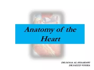
Anatomy of the Heart. The heart is located in the chest cavity, surrounded by the pericardial sac, in the anterior portion of the mediastinum. . The Pericardium. Pericardial cavity. The pericardium is a double-walled sac (pericardial sac) that encloses the heart. Parietal pericardium.

E N D
The heart is located in the chest cavity, surrounded by the pericardial sac, in the anterior portion of the mediastinum.
The Pericardium Pericardial cavity The pericardium is a double-walled sac (pericardial sac) that encloses the heart. Parietal pericardium Visceral pericardium (epicardium) Mesothelium Pericarditis is a disorder caused by inflammation of the pericardium, the sac-like covering of the heart. Areolar tissue Fibrous tissue Pericarditis can be caused by bacterial, fungal, or viral infections. It may also be a result of injury or trauma to the chest, esophagus, or heart. Pain occurs as a result of the inflamed pericardium rubbing against the heart.
The Heart Wall Parietal pericardium Areolar tissue Areolar tissue Pericardial cavity MYOCARDIUM (cardiac muscle tissue) ENDOCARDIUM EPICARDIUM Endothelium Mesothelium Visceral pericardium Mesothelium Areolar tissue Fibrous tissue Endocarditis is inflammation of the inside lining of the heart chambers and heart valves (endocardium). Most people who develop endocarditis have heart disease of the valves.
An Introduction to the Cardiovascular System GasExchange Systemic Pulmonary Circuit Circuit Capillary Lung Venule Arteriole Pulmonary arteries Pulmonary veins O2 poor, CO2 rich blood O2 rich, CO2 poor blood Wastes Nutrients O2 CO2 CO2 O2 Venae cavae Aorta Capillary Tissue Venule Arteriole
Cardiovascular System: Pulmonary Circuit It carries blood to the lungs for gas exchange and returns it to the heart. Systemic Circuit It supplies blood to every organ of the body, including the lungs and the heart itself. Blood Vessels: Arteries They carry blood away from the heart. Veins They carry blood back to (toward) the heart. Capillaries They connect the arteries with the veins.
Gas exchange Pulmonary Circuit It carries blood to the lungs for gas exchange and returns it to the heart. O2rich blood through VEINS O2poor blood through ARTERIES Systemic Circuit It supplies blood to every organ of the body, including the heart itself. O2rich blood through ARTERIES O2poor blood through VEINS
Internal Anatomy and Organization Gas exchange Poor oxygen blood Pulmonary Arteries (2) Reach oxygen blood Superior vena cava Coronary sinus Inferior vena cava Pulmonary veins (4) Pulmonary Trunk RIGHT ATRIUM LEFT ATRIUM To the rest of the body RIGHT VENTRICLE LEFT VENTRICLE Aorta
Superior vena cava It drains oxygen-poor blood from tissues and organs superior to the diaphragm to the right atrium. Aorta Pulmonary trunk It carries oxygen-rich blood from the left ventricle to the whole body. It carries oxygen-poor blood from the right ventricle to the lungs. Pulmonary veins (4) Inferior vena cava They carry oxygen-rich blood from the lungs to the left atrium. It drains oxygen-poor blood from tissues and organs inferior to the diaphragm to the right atrium. Coronary sinus (no shown) It drains oxygen-poor blood from the heart tissues to the right atrium.
The Heart Valves The heart has two pairs of one-way valves that prevent the backflow when the chambers contract It prevents back flow of blood from the pulmonary trunk to the RV Aortic semilunar valve It prevents back flow of blood from the LV to the LA It prevents back flow of blood from the RV to the RA Left AV (bicuspid) valve Pulmonary semilunar valve It prevents back flow of blood from the aorta to the LV Right AV (tricuspid) valve
Heart Sounds During ventricular systole (contraction) the two AV close at the same time and produce the first sound referred asLubb. Lubb Dupp Dupp Lubb When the ventricles relax (diastole) the two semilunar valves close at the same time and produce the second sound referred asDubb.
Heart Sounds The heart sounds are described as a lubb-dupp sound First Sound ( “lubb” ): It is the strongest one. It is produced by the closing of the AV valves Second Sound ( “dupp” ):It is produced by the closing of the semilunar valves Normal valves produce high pitch sound Incompetent valves (that do not close completely) produce a switching sound as blood flows back creating abnormal sounds Heart Murmur: It is a sound produced by regurgitation through valves
Ligamentum arteriosum Four openings of the pulmonary veins Remnant of ductus arteriosum Opening of superior vena cava Aortic arch (Remnant of foramen oval) Pulmonary trunk Fossaovalis Pulmonary veins Opening of coronary sinus Aortic semilunar valve Opening of inferior vena cava It prevents back flow of blood from the aorta to the LV Left AV valve or bicuspid valve Right AV valve or tricuspid valve It prevents back flow of blood from the RV to the RA (mitral valve) It prevents back flow of blood from the LV to the LA Pulmonary semilunar valves It prevents back flow of blood from the pulmonary trunk to the RV
Cusps Chordae tendineae Papillary muscle Trabeculae carneae Transverse section, superior view
Circumflex artery Left coronary artery Anterior I-V artery Posterior I-V artery Right coronary artery Right coronary artery Marginal arteries Left coronary artery Coronary sinus Great cardiac vein Circumflex artery Anterior interventricular artery Posterior interventricular artery Small cardiac vein Marginal artery Middle cardiac vein The Blood Supply to the Heart The Coronary Arteries
Atrioventricular bundle or bundle of His Right and left bundle branches Sinoatrial node (SA node) Atrioventricular node (AV node) Purkinje fibers The Conducting System It connects electrically the atria to the ventricles. They conduct the impulse to the Purkinje fibers. It establishes the heart rate (pacemaker). It delays the impulses to allow the atria to finish contracting before the ventricles start to contract. They conduct the impulse to the lateral walls of the ventricles allowing the contraction to spread from the apex to the base.
Impulse Conduction through the Heart SA node fires and atrial activation begin. Time 0 5 2 3 4 5 5 1 2 3 4 1 Stimulus spreads across the atrial surfaces and reaches the AV node. Elapsed time: 50 msec There is a 100 msec delay at the AV node. Atrial contraction begins. AV node fires. Elapsed time : 150 msec The impulse travels along the inter-ventricular septum within the AV bundle and the bundle branches to the Purkinje fibers. Elapsed time: 175 msec The impulse is distributed by Purkinje fibers and relayed through the ventricular myocardium. Atrial contraction is completed and ventricular contraction begins. Elapsed time: 225 msec
The Cardiac Cycle At the beginning of their contraction (systole) the ventricles contracts isovolumetrically (the pressure increases but the volume inside the ventricles does not changes). In the period of isovolumetric contraction, the ventricles contract and the pressure rises, but blood does not flow because all the valves are closed. Pressure Pressure
The Cardiac Cycle At the beginning of their contraction (systole) the ventricles contracts isovolumetrically (the pressure increases but the volume inside the ventricles does not changes). In the period of isovolumetric contraction, the ventricles contract and the pressure rises, but blood does not flow because all the valves are closed. Pressure Pressure Once pressure in the ventricles exceeds that in the arterial trunks (pulmonary and aortic), the semilunar valves open and blood flows into the pulmonary and aortic trunks. This point marks the beginning of the period of ventricular ejection.
(b) At the start of the atrial systole, the ventricles are already filled to about 70% of their normal capacity, due to passive blood flow. At the end of the atrial systole, each ventricle contains a maximum amount of130 mL of blood: End-diastolic volume (a) (c) In the period of isovolumetric contraction, the ventricles contract and the pressure rises, but blood does not flow because all the valves are closed. (d) (e) This point marks the beginning of the period of ventricular ejection.
(f) At the start of the atrial systole, the ventricles are already filled to about 70% of their normal capacity, due to passive blood flow. (a) Atrial Systole A small amount of blood (30 %) is forced to the ventricles Ventricular contraction closes the AV valves (first sound). Isometric contraction. Fist Phase: Ventricular Systole Pressure increases and semilunar valves open. Ventricular ejection. Second Phase: Pressure decreases in the ventricles and semilunar valves close (second sound). Early: Ventricular Diastole Atria are also in diastole. Passive blood flow fills the ventricles (70%). Late: Atrial Diastole Ventricles are also in diastole. Passive blood flow fills the ventricles (70%).
Electrocardiogram or ECG (EKG) It is the graphic recording of the electrical activity of the heart as it works
The Electrocardiogram R QRS complex +1 Depolarization of ventricles Depolarization of atria T P Repolarization of ventricles 0 Q S PQ segment ST segment It represents the time during which the ventricles contract and eject blood Millivolts
0.8 sec 0.5 sec 75 bpm Sinus Rhythm (normal) 120 bpm Tachycardia 1.4sec 1.4sec 0.5 sec 0.3 sec 1.4sec 46 bpm Bradycardia Arrhythmia Extrasystole Nodal Rhythm Ventricular fibrillation Heart block
R P T Q S
QRS complex Depolarization of ventricles Depolarization of atria Repolarization of ventricles T P ST segment ATRIAL ATRIAL ATRIAL DIASTOLE VENTRICULAR VENTRICULAR VENTRICULAR SISTOLE DIASTOLE SISTOLE DIASTOLE DIASTOLE
End-Diastolic Volume (EDV) It is the volume of blood that each ventricle contains at the end of ventricular filling (about 130 mL). Stroke Volume (SV) It is the volume of blood that each ventricle ejects during ventricular ejection (about 70 - 80 mL). End-Systolic Volume (ESV) It is the volume of blood left behind in the ventricles after ventricular ejection. EDV – SV = (ESV) Ejection fraction It is the percentage of the end-diastolic volume (EDV) that is ejected (about 54%). Cardiac Output (CO) The amount of blood pumped by the left ventricle in one minute Cardiac Output (CO) = Stroke Volume (SV) x Heart Rate (HR) 75 bpm x 80 mL/beat = 6000 mL/min (6L/min)
Superior vena cava (to the right atrium) Aortic arch Ascending aorta S A node (pace maker) (from the left ventricle) Right auricle Left auricle (from the right ventricle) Pulmonary trunk Left coronary artery Right ventricle Left ventricle Right coronary artery Circumflex artery Anterior interventricular artery Marginal artery Anterior view
(to the left atrium) Right pulmonary veins Right auricle Left pulmonary veins Left auricle (to the left atrium) (to the right atrium) Inferior vena cava Great cardiac vein Right ventricle Left ventricle Coronary sinus Posterior cardiac vein Small cardiac vein Middle cardiac vein Posterior interventricular artery (branch of the right coronary artery) Posterior view
Internal Structures Superior vena cava Aortic arch Conducting system Ligamentum arteriosum S A node (pace maker) Pulmonary trunk Ascending aorta Orifices of coronary arteries Aortic semilunar valve AV node Right AV valve or tricuspid valve Left AV valve or bicuspid valve Chordae tendineae Atrioventricular bundle (of His) Papillary muscles Left bundle branch Right bundle branch Trabeculae carneae Purkinje fibers