Download
1 / 40
420 likes | 729 Vues
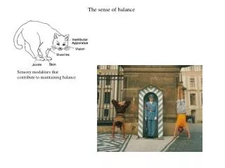
The sense of balance. Sensory modalities that contribute to maintaining balance. The vestibular system (labyrinth).

E N D
The sense of balance Sensory modalities that contribute to maintaining balance
The vestibular system (labyrinth) The inner ear is divided into vestibular division, which contains the organs of equilibrium (the utricle, saccule, and semicircular canals) and cochlear division, which contains the organs of hearing (the cochlea).
The vestibular system (labyrinth) The otolith organ are detecting linear acceleration. It consists of two sacs and the receptors are grouped in a macula in each sac. Three semicircular canals are detecting rotation (angular acceleration). Each semicircural canal has an enlargement (ampulla) within which the sensory cells are grouped.
Hair cells Hair cells are receptors responsible for the sense of equilibrium and for audition. The hair cells of the receptor organs in the human internal ear are of similar form and function. They transduce mechanical stimuli into receptor potential. This scanning electron micrograph of a hair cell's apical surface. The hair bundle comprises some 60 stereocilia, arranged in rows of varying length and the single kinocilium at the bundle's tall edge. Deflection of the hair bundle to the right, the positive stimulus direction, depolarizes the hair cell; movement in the opposite direction elicits a hyperpolarization. From the cell's apical surface extends the hair bundle, the mechanically sensitive organelle. Afferent and efferent synapses occur upon the basolateral surface of the plasma membrane.
Tip links Stereocilia are connected with each other by elastic structures within the hair bundle called tip links. The scanning electron micrograph of a hair bundle's top surface. The links that connect each stereociliary tip to the side of the longest adjacent stereocilium are visible.
A model for the mechanism of mechanoelectrical transduction by hair cells The ion channels that participate in mechanoelectrical transduction in hair cells are gated by elastic structures in the hair bundle. Opening and closing of these channels is controlled by the tension in the gating spring, that senses hair-bundle displacement. The membrane potential of the receptor cell depends on the direction in which the hair bundle is bent. Deflection toward the kinocilium causes the cell to depolarize and thus increases the rate of firing in the afferent fiber. Bending away from the kinocilium causes the cell to hyperpolarize, thus decreasing the afferent firing rate.
Adaptation in the hair cells The electrical response to a positive stimulus displays an initial depolarization, followed by a decline to a plateau and an undershoot at the cessation of the stimulus. Negative stimulation elicits a complementary response. Bundle movement in response to positive stimulation increases tip link tension and opens transduction channels. As stimulation continues, the tip link's upper attachment moves down the stereocilium, allowing each channel to close during adaptation. During negative stimulation tension is restored to the initially slack tip link by active ascent of the link's upper insertion.
Mechanism of transduction • Movement • Streching of the gating string • Increase membrane conductance to K+ • Influx of K+ ions into the cell, the depolarization spreads into the cell • Release of neurotransmitter at the hair cell synapse onto vestibular nerve sensory terminal.
The Otolith Organs In the otolith organs, the sensory epithelium is overlain with a sheet of gelatinous extracellular matrix, the otholitic membranne. Embedded in this structure are otoconia, rocklike cristals of calcium carbonate. When the head undergoes linear acceleration the otoconial mass lags behind movement of the head, because of its inertia. The motion of the otoconia is communicated to the otolithic membrane, which thus shifts with respect to the underlying epithelium. This motion in turn deflects the hair thus exciting an electrical response in the hair cells. Otholitic membranne
Spatial organization of the otolith organs The hair cells in each organ are localized to a roughly elliptical patch, called the macula. The hair bundles are oriented orthogonally to a curving midline (the striola). This arrangement allows for detection of all possible stimulus orientations within the plane of the macula. Any particular horizontal acceleration maximally depolarizes one group of hair cells and maximally inhibits a complementary set. Because the organs are bilateral, the brain receives additional information from the contralateral labyrinth. The utricles are oriented horizontally, the saccules are oriented vertically. The hair cells in the utricles respond to accelerations in the horizontal plane; saccules are especially sensitive to vertical accelerations, of which gravity is the most important.
The semicircular canals A thickened zone of epithelium, the ampula, contains the hair cells. The hair bundles of the hair cells extend into a gelatinous cupula, which stretches from the crista to the roof of the ampulla. The cupula is displaced by the flow of endolymph when the head moves. As a result, the hair bundles extending into the cupula are also displaced. Because all the hair bundles in each semicircular canal share a common orientation, angular acceleration in one direction depolarizes hair cells while acceleration in the opposite direction hyperpolarizes the receptor cells and diminishes spontaneous neural activity.
Orientation of the semicircular canals In each labyrinth the three canals are almost precisely perpendicular to one another, so that the canals represent accelerations about three mutually orthogonal axes. The planes in which the semicircular canals lie do not, however, correspond with the head's major anatomical planes.
Menière Disease • Menière disease affects the vestibular labyrinth. • It is characterized by relapsing vertigo (dizziness), with attacks lasting from tens of minutes to tens of hours. The vestibular symptoms are often accompanied by noise in the ears (tinnitus) and distorted hearing. • It is believed to be linked to an excess of endolymph in the inner ear. • Removal of the affected labyrinth surgically may relieve severe vertigo. • Vincent Van Gogh may have suffered from Menière disease. Vincent van Gogh, Selfportrait, 1889 K. Arenberg, L. F. Countryman, L. H. Bernstein and G. E. Shambaugh Jr,Van Gogh had Meniere's disease and not epilepsy, JAMA,Vol. 264 No. 4, July 25, 1990 Arnold, Wilfred N. (1992). "Vincent van Gogh: Chemicals, Crises, and Creativity". Birkhäuser Boston.
Vestibular pathways The axons that carry the hair cells responses to the central nervous system terminate in several large cell groups (vestibular nuclei) in the brainstem. From there the signals go to neck and trunk motoneurons, to limb motoneurons, to cerebellum, thalamus and extraocular motoneurons. This network of vestibular connections is responsible for the various reflexes that the body uses to compensate for head movement and the perception of motion in space.
The vestibulo-ocular reflex Rotation of the head in a counterclockwise direction causes endolymph to move clockwise with respect to the canals. This reflects the stereocilia in the left canal in the excitatory direction, thereby exciting the afferent fibers on this side. In the right canal the hair cells are hyperpolarized and afferent firing there decreases. The vestibulo-ocular keeps the visual images fixed on the retina, by moving the eyes when the head moves. Vestibular nerve signals head velocity to the vestibular nuclei and motoneurons that control ocular muscles. E.g., Counterclockwise head rotation excites the left horizontal canal, which then excites neurons that evoke rightward eye movement.
Weightlessness • Pleasant and intriguing experience • Space adaptation syndrome (SAS) – vomiting, headache, nausea • Fast adaptation (< 72 hours) • Upon return to Earth after long-term weightlessness, slow adaptation, e.g., in maintaining standing balance with eyes closed.
Sounds Hearing ranges of different animals
Outer ear The ear has three functional parts. The main part of the external ear, the auricle the auricle captures sound efficiently sends it into the ear canal. Our capacity to localize sounds in space, especially along the vertical axis, depends critically upon the sound - gathering properties of the external ear.
Middle ear Middle ear transmits mechanical energy to the ear's receptive organ. The three tiny ossicles, or bones: the malleus, or hammer; the incus, or anvil; and the stapes, or stirrup pass the motions of the tympanic membrane to the oval window – the bony covering of the cochlea.
Impedance matching water– inner ear air – environment Amplitude reflection and transmission coefficients Energy reflection and transmission coefficients Mechanical impedance Mass density Wave propagation speed
Impedance matching Energy reflection and transmission coefficients: fraction of power transmitted ~99.9% is reflected If only 1 part in 1000 makes it thru, the loss is: 10log101/1000 = -30 dB
Transformer Action #1 Incus Malleus Stapes ~ 3.2 mm2 Tympanic membrane ~ 55 mm2 Area ratio about 17:1 Pressure = Force/area Stapes pressure is 17 times TM Sound pressurelevelis 20log10P/Pref Sound pressure increase is approximately 20log1017/1 = 25dB
Transformer Action #2 Incus 7 mm Malleus 9 mm F1 x L1 = F2 x L2 Malleus/Incus length ratio is 9:7 soforce and hence the pressure is increased by 1.3:1 Sound pressure increase is approximately 20log109/7 = 2 dB
Eardrum displacement at threshold of hearing Acoustic travelling wave Total wave energy (Epot = Ekin) Wave intensity Wave intensity at the threshold of hearing 0 dB Wave amplitude Wave amplitude at 440 Hz Hydrogen atom diameter The eardrum displacement at threshold of hearing is ~ 1/10 the diameter of a hydrogen atom!
Sound Intensity and Sound Pressure Levels Human ear responds to sound intensities in the range of 10-12– 100 W/m2. Sound intensity level is defined as: • where: • – sound intensity level I – sound intensity I0 – standard reference sound intensity 10–12 W/m2 Sound pressure level (SPL) or sound level is a logarithmic measure of the effective sound pressure of a sound relative to a reference value:
Sound Intensity and Sound Pressure Levels Intensity dB Pressure Examples 108 W/m2 200 2*105 Pa Volcano erruption 102 W/m2 140 2*102 Pa Jet aircraft, 50 m 1W/m2 120 2*101 Pa Rock concert 10-2W/m2 100 2 Pa Disco, 1m from the speaker 10-4 W/m280 2*10-1 Pa Highway, 5 m 10-6 W/m260 2*10-2 Pa Speech, 1 m 10-8 W/m240 2*10-3 Pa Background in quiet library 10-10 W/m220 2*10-4Pa Background in TV studio 10-12 W/m20 2*10-5Pa Hearing threshold Threshold of pain 130 dB Discomfort threshold, hearing damage possible 120 dB limit Apple iPod volume in Europe 85 dB
How cochlea works ? High frequency Low frequency Cochlea in the inner ear is shaped like a snail shell. Net effect is to boost pressure created by sound so that the inner ear fluid can be displaced. Its spiral shape effectively boosts the strength of the vibrations caused by sound, especially for low frequencies.
Cochlea and basilar membrane Cochlea contains basilar membrane, which separates two liquid-filled tubes that run along the cochlea, the scala media and the scala tympani. Basilar membrane is narrow at the base and widens at the tip. Different frequencies are coded by the position along the membrane – high frequencies displace the membrane at the base, low frequencies displace the membrane at the apex. The organ of Corti sits on top of the basilar membrane. It transduces pressure waves to action potentials.
Helmholtz’s resonance theory of hearing Different frequencies of sound are encoded by their precise position along the basila membrane. Short strings at the base would resonate in response to high notes, and the long strings (at the apex) to low notes.
The travelling wave theory - Von Bekesy (1928). Nobel 1961 The sound pressure applied to the oval window is transmitted as a travelling wave along the basila membrane. The peak diplacements for high frequencies are toward the base, and for low frequencies are toward the apex. Georg von Békésy(1899 –1972) Envelopes induced by sound at 3 different frequencies
Problem: envelopes of the travelling waves are wide while we are hearing pure tones There must be additional mechanism for tunning of the auditory system to the sound frequency. Proof: movements of the basilar membrane Effect of cochlear amplifier. C) The peak due to cochlear amplifier. D) Amplitude of the passive movement of basilar membrane in the absence of the cochlear amplifier.