Download
1 / 162
1.77k likes | 2.69k Vues
The Sense of Hearing. Prof.Dr. Ümmühan İşoğlu-Alkaç YU, 03.03.2014. References:. Textbook of Medical Physiology, Guyton & Hall Lippincot’s Illustrated Reviews Physiology Preston & Wilson. Ear. Structure The auditory system, or ear, can be divided into three main anatomical components:

E N D
The Sense of Hearing Prof.Dr. Ümmühan İşoğlu-Alkaç YU, 03.03.2014
References: • Textbook of Medical Physiology, • Guyton & Hall • Lippincot’s Illustrated Reviews Physiology • Preston & Wilson
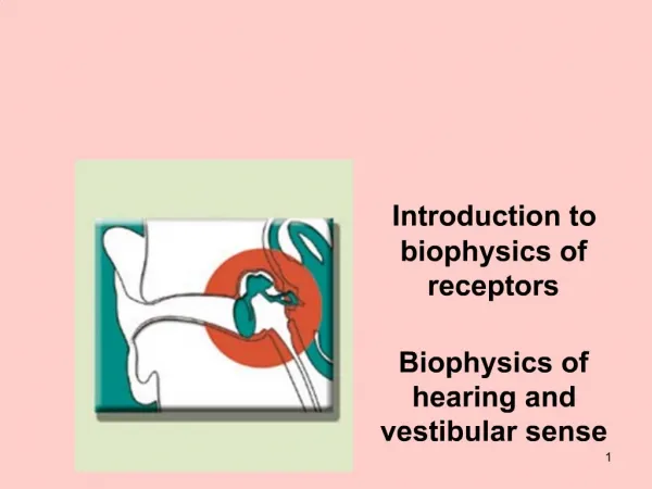
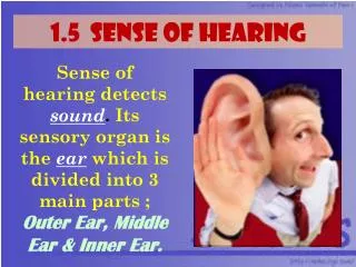
Ear Structure The auditory system, or ear, can be divided into three main anatomical components: • the outer, • middle, and • inner ear
Outer ear: The pinna collects and focuses sounds. Sounds arechanneled into the ear canal (external auditory meatus), which allows them to pass through the skull’s temporal bone. The canalends blindly at the tympanic membrane (eardrum), which vibrates in response to sound.
Middle ear: The middle ear is an air-filled chamber lying betweenthe eardrum and the inner ear. It connects with the nasopharynxvia the eustachian tube, which drains fluids and allows thepressure across the eardrum’s two surfaces to equalize. Eardrumvibrations are transmitted to the inner ear by an articulating leversystem comprising three, small, fragile bones called ossicles.
The bones are known as the malleus (hammer), incus (anvil), and stapes (stirrup), their names roughly reflectingtheir shapes. The malleus is attached to the eardrum’s innersurface and transmits vibrations to the incus. The incus transmitsthem to the stapes. The stapes’ footplate inserts into and is firmlyattached to the oval window of the inner ear.
Inner ear: The inner ear contains a convoluted series of fluid-filledchambers and tubes (membranous labyrinth). The structuresare encased within bone (the bony labyrinth) with a thin layerof perilymph trapped between bone and membranes. The labyrinthhas two sensory functions.
The auditory portion is calledthe cochlea. The vestibular portion contributes to our sense ofbalance. It comprises the otolith organs (utricle and saccule) and three semicircular canals.
Tympanic membrane (eardrum) Middleear Malleus İncus Stapes Innerear Cochlea
Kulak anatomik olarak üç bölgeden meydana gelir. Dış kulak yolu kulak kepçesinden başlar ve timpan zarına kadar devam eder. Orta kulak içinde üç adet kemikçik yer alır. İç kulaktaki birbiri ile bağlantılı iki yapıdan sadece kohlea işitme ile ilgilidir. Yarım daire kanalları ise vestibüler sistem ile ilişkilidir.
Kulak anatomik olarak üç bölgeden meydana gelir. Dış kulak yolu kulak kepçesinden başlar ve timpan zarına kadar devam eder. Orta kulak içinde üç adet kemikçik yer alır. İç kulaktaki birbiri ile bağlantılı iki yapıdan sadece kohlea işitme ile ilgilidir. Yarım daire kanalları ise vestibüler sistem ile ilişkilidir.
Kohleanın iç kısmı üç bölmeden oluşur. Scala media ve scala tympani’yi ayıran baziller membran üzerinde yer alan Corti organı, ses dalgalarının sinir sinyallerine dönüştüğü reseptör organdır.
Timpan zarını titreştiren ses dalgasının orta kulak kemikçiklerinden oval pencere aracılığıyla kohleaya iletilmesinden sonra baziller membran üzerinde yaptığı etki. Şekilde etkinin daha iyi gösterilebilmesi için, kendi üzerinde kıvrımlar oluşturan kohlea düz bir boru gibi gösterilmiştir.
Tympanic membrane, ossicular system of the middle ear, and inner ear
Impedance Matching The inner ear is filled with fluid that has a high inertia and is difficultto move compared with air. The middle ear’s function is, thus, to harnessthe sound wave’s inherent energy and transmit it to the inner earwith sufficient force to overcome the inertia of the fluid contents. Thisprocess is called impedance matching.
Mechanism: The ossicles form a lever system that amplifieseardrum movements by 30%. It also focusesthe movements on the stapes’ footplate, whose surface area is17 times smaller than the eardrum. Amplification and focusingcombined increase force per unit area 22-fold, which allowssounds to be transferred to the inner ear with sufficient force toovercome cochlear fluid inertia.
Because fluid has far greater inertia than air does, it is easily understoodthat increased amounts of force are needed to cause vibration in the fluid. Therefore, the tympanic membrane and ossicular system provide impedancematching between the sound waves in air and the sound vibrations in the fluidof the cochlea. Indeed, the impedance matching is about 50 to 75 per cent ofperfect for sound frequencies between 300 and 3000 cycles per second, whichallows utilization of most of the energy in the incoming sound waves.
In the absence of the ossicular system and tympanicmembrane, sound waves can still travel directlythrough the air of the middle ear and enter the cochleaat the oval window. However, the sensitivity forhearing is then 15 to 20 decibels less than for ossiculartransmission—equivalent to a decrease from amedium to a barely perceptible voice level.
Attenuation of Sound by Contraction of the Tensor Tympani andStapedius Muscles. When loud sounds are transmittedthrough the ossicular system and from there into the central nervous system, a reflex occurs after a latent period of only 40 to 80 milliseconds to cause contractionof the stapedius muscle and, to a lesser extent, thetensor tympani muscle.
The tensor tympani muscle pulls the handle of the malleus inward while thestapedius muscle pulls the stapes outward. These twoforces oppose each other and thereby cause the entireossicular system to develop increased rigidity, thusgreatly reducing the ossicular conduction of lowfrequencysound, mainly frequencies below 1000 cycles per second.
This attenuation reflex can reduce the intensity oflower-frequency sound transmission by 30 to 40 decibels,which is about the same difference as thatbetween a loud voice and a whisper.
The function ofthis mechanism is believed: 1. To protect the cochlea from damaging vibrationscaused by excessively loud sound. 2. To mask low-frequency sounds in loud environments. This usually removesa major shareof the background noise and allows a person toconcentrate on sounds (above 1000 cycles/sec), where most of the pertinent informationin voice communication is transmitted. 3. To decrease a person’s hearingsensitivity to his or her own speech.
Damping: Lever system flexibility is modulated to reduce soundamplitude under certain circumstances. The malleus and stapesare attached to two tiny muscles under autonomic control. The tensor tympani anchors the malleus to thewall of the middle ear and is innervated by the trigeminal nerve (cranial nerve [CN] V).
The stapes is anchored by the stapedius, which is innervated by the facial nerve (CN VII). When the twomuscles contract, the ossicular chain becomes more rigid, and sound transmission is attenuated. The attenuation reflex can betriggered by loud sounds but is probably designed to dampen thesound of our own voices when talking.
Functional Anatomy of the Cochlea The consists of three tubes coiled side by side: (1)the scala vestibuli, (2) the scala media, and (3) the scala tympani. The scala vestibuli and scala media are separated from each other by Reissner’s membrane (vestibular membrane), the scala tympani and scala media are separated fromeach other by the basilar membrane.
On the surface ofthe basilar membrane lies the organ of Corti, whichcontains a series of electromechanically sensitivecells, the hair cells. They are the receptive end organsthat generate nerve impulses in response to sound vibrations.
Reissner’s membrane is so thin and so easilymoved that it does not obstruct the passage of soundvibrations from the scala vestibuli into the scala media. Therefore, as far as fluid conduction of sound is concerned,the scala vestibuli and scala media are consideredto be a single chamber
Cochlea The cochlea is a long (3 cm), tapered tube containing three fluidfilled chambers that run the length of the tube. The tube is coiledlike a snail shell in vivo, but the functional architecture is easier to understand when considered uncoiled. The three chambersare called the scala vestibuli, the scala media, and the scala tympani.
Scala vestibuli and scala tympani: The upper and lower chambers are both filled with perilymph (a fluid approximating plasma) and are physically connected by a small opening (the helicotrema)at the cochlear apex.
Scala media: The center chamber is separated from the scalavestibuli by the Reissner membrane (or vestibular membrane)and from the scala tympani by the basilar membrane. It is filled with endolymph, a K-rich fluid produced by the stria vascularis, a specialized epitheliumlining one wall of the chamber. The scalamedia contains the organ of Corti, which is the auditory sensoryorgan.
AUDITORY TRANSDUCTION Sound waves enter the cochlea via the oval window, which forms thebasal end of the scala vestibuli. Stapes motion sets up apressure wave in the perilymph that runs down the chamber’s length tothe apex, passes through the helicotrema, and then pulses back down the scala tympani to the cochlear base. Here, it encounters the roundwindow, a thin membrane located between the inner and middle ear.
Movement of fluid in the cochlea after forward thrust of the stapes.
The membrane vibrates back and forth in reverse phase with the wavegenerated by stapes movement. The stapes would not be able to displacethe oval window and set the perilymph in motion if the round windowdid not exist because the cochlear chamber walls are otherwise rigidlyencased in bone. The scala media, which approximates a fluid-filledsac suspended between the two chambers, is buffeted by the pressurewave as it pulses back and forth.
Thus, although the sound wave never enters the scala media directly, the entire structure wobbles, much as awaterbed responds when pushed down on hard at one corner. It is thisbuffeting that is sensed by the organ of Corti.
Basilar Membrane and Resonance in the Cochlea. Thebasilar membrane is a fibrous membrane that separates the scala media from the scala tympani. It contains 20,000 to 30,000 basilar fibers that projectfrom the bony center of the cochlea, the modiolus, toward the outer wall.
These fibers are stiff, elastic,reedlike structures that are fixed at their basal endsin the central bony structure of the cochlea (the modiolus)but are not fixed at their distal ends, exceptthat the distal ends are embedded in the loose basilarmembrane. Because the fibers are stiff and free at one end, they can vibrate like the reeds of a harmonica.
The lengths of the basilar fibers increase progressivelybeginning at the oval window and going fromthe base of the cochlea to the apex, increasing from alength of about 0.04 millimeter near the oval andround windows to 0.5 millimeter at the tip of thecochlea (the “helicotrema”), a 12-fold increase in length.