Download
1 / 24
250 likes | 572 Vues
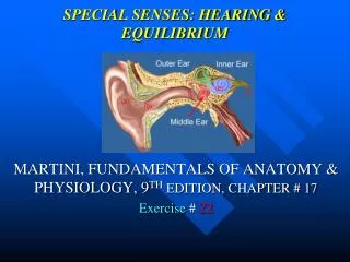
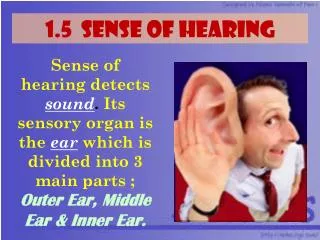
Sense of Hearing and Equilibrium. Coach O’Rourke Brazos HS. 3 Parts. Sense of Hearing Made up of: Outer ear Middle ear Inner ear Ear also functions as sense of equilibrium. Outer (External) Ear. 3 parts Auricle (Pinna) Funnel-like structure

E N D
Sense of Hearing and Equilibrium Coach O’Rourke Brazos HS
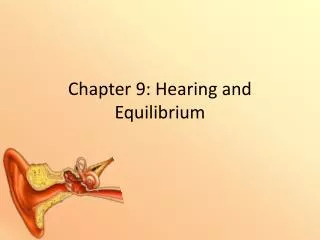
3 Parts • Sense of Hearing • Made up of: • Outer ear • Middle ear • Inner ear • Ear also functions as sense of equilibrium
Outer (External) Ear • 3 parts • Auricle (Pinna) • Funnel-like structure • External Acoustic Meatus (External Auditory Canal) • S-shaped tube • Eardrum (tympanic membrane) • Cone shaped • Sometimes considered part of middle ear
Auricle • Helps collect sound waves traveling through the air and directs them into the external acoustic meatus.
External Acoustic Meatus • As sound waves enter they change the pressure on the eardrum.
Eardrum • Semitransparent membrane covered by a thin layer of skin on its outer surface and a mucosa membrane in the inside • Has an oval margin and cone-shape with the cone apex pointing inward.
Middle Ear (Tympanic Cavity • Air-filled space in the temporal bone • Contains three bones called Auditory Ossicles • Malleus • Incus • Stapes • Oval Window • Opening of tympanic cavity that leads to inner ear • Auditory Tube (Eustachian Tube) • Connects middle ear to nasal cavity
Auditory Ossicles • Tiny ligaments attach bones to wall of tympanic cavity • Are covered by mucous membrane • Bridge the ear drum and the inner ear • Transmit vibrations between eardrum and I.E. • Malleus is attached to ear drum • Once eardrum vibrates so does malleus which causes Incus and then Stapes to vibrate in unison. • Stapes is attached to oval window • Help increase(amplify) the force of vibrations from eardrum to oval window • Oval window vibrations move fluid in inner ear to stimulate hearing receptors
Auditory TubeEustachian Tube • Connects middle ear to back of nasopharynx • Conducts air between tympanic cavity and the outside of body by way of nose and mouth • Helps maintain equal pressure of both sides of eardrum • Function is noticeable during rapid altitude changes • Popping sound is heard when hearing is restored back to normal
Inner (Internal) Ear • Consists of: • Labyrinth • Semicircular canals • Cochlea • Round window • Spiral Organ (Organ of Corti)
Labyrinth • Complex system of communicating chambers • 2 Parts: • Osseous Labyrinth • Bony canal in temporal bone • Membranous Labyrinth • Similar shape of osseous labyrinth • Found within osseous labyrintjh • Fluid between labyrinths is called perilymph which is secreted by cells in the walls of the bony canal • Fluid within Membranous is called endolymph
Semicircular Canals and Cochlea • SCC • Part of labyrinth • Provide sense of equilibrium • Cochlea • Functions in hearing • Has bony core and thin bony shelf that winds around the core like the threads of a screw • Shelf divides the osseous labyrinth of cochlea into upper and lower compartments • Upper Compartment (ScalaVestibuli) • Leads from oval window to apex of spiral • Lower Compartment (Scala Tympani) • Extends from Apex to round window
Round Window • Opening in the wall of inner ear • Cochlear Duct • Part of membranous labyrinth • Lies between to bony compartments and ends as a closed sac at the apex • Separated from scalavestibuli by vestibular membrane (Reissner’s membrane) • Separated from Scala Tympani by basilar membrane Cochlea Cont.
Spiral Organ • AKA Organ of Corti • Contains hearing receptors • Receptor Cells • AKA Hair cells • Function somewhat like neurons • Move back and forth depending on pitch of sound • Young person • Detect sound waves ranging from 20-20,000 or more vibrations per second • 2,000-3,000 is the range of greatest sensitivity
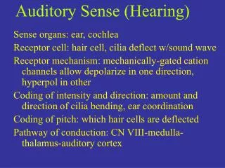
Nerve Pathways • Temporal Lobes interpret hearing • Follow the Vestibulocochlear cranial nerve
Sense of Equilibrium • 2 Senses-2 different sensory organs • Static • Sense position of head • Maintain stability and posture when head is still • Dynamic • Sudden movement or rotation of head • Aid in maintaining balance
Static Equilibrium • Vestibule • Bony chamber between SCC and Cochlea • Contains: • Utricle • Saccule • Each of these have tiny hair like structures called macula • Head bending forward, backward, or to one side stimulate hair cells
Dynamic Equilibrium • Organs are the three SCC • Contain: • Ampulla • Houses sensory organs called crista ampularis contain a number of sensory hair cells and supporting cells • Rapid turns of head stimulate crista ampularis • SCC move with head but fluid stays stationary • Cerebellum • Parts interpret impulses from SCC • Other organs help maintain balance • Eyes • Joints in neck