Altered Hematologic Function part 2
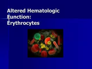
Altered Hematologic Function part 2. Alterations in Leukocytes and Blood Coagulation. Leukocytes. White blood cells Defend body through: the inflammatory process phagocytosis removal of cell debris immune reactions. White Blood Cell Types: Granulocytes and Agranulocytes.

Altered Hematologic Function part 2
E N D
Presentation Transcript
Leukocytes • White blood cells • Defend body through: • the inflammatory process • phagocytosis • removal of cell debris • immune reactions
White Blood Cell Types:Granulocytes and Agranulocytes • Granulocytes –visible granules in the cytoplasm. • Granules contain: • Enzymes • Other biochemicals that serve as signals and mediators of the inflammatory response
Granulocyte cell types: • Neutrophils – phagocytes • Eosinophils – red granules, associated with allergic response and parasitic worms • Basophils – deep blue granules - Release heparin, histamine and serotonin
Agranulocytes • Granules too small to be visible • Monocytes – become macrophages • Lymphocytes – B cells and T cells = immune functions
WBC’s originate in red bone marrow from stem cells. • Granulocytes mature in the marrow and have a lifespan of hours to days • Agranulocytes finish maturing in blood, or in other locations. Monocytes live about 2 - 3 months, lymphocytes for years.
Types of stem cells: • Pluripotent • Multipotent • Committed progenitor cells • Multipotent blood cells: • Common lymphoid • Common myeloid • Committed stem cell makes specific blood cells (CFU) – stimulated by erythropoietin, thrombopoietin, granulocyte-mononcyte CSF
Production of WBC’s increases in response to : • Infection • Presence of steroids • Decreased reserve of leukocyte pool in bone marrow
WBC Abnormalities • Leukocytosis – increased numbers of WBC’s • May be a normal protective response to physiological stressors • Or may signify a disease state – a malignancy or hematologic disorder • Leukopenia – decreased numbers of WBC’s – this is never normal • Increases the risk of infections. • Agranulocytosis = granulocytopenia
Leukeopenia may be due to: • Radiation • Anaphylactic shock • Autoimmune disease • Chemotherapeutic agents • Idiosyncratic drug reactions • Splenomegaly • infections
Mononucleosis • Self-limiting lymphoproliferative disorder caused by the Epstein-Barr Virus • Infects 90% of people • Incorporates into DNA of B cells causing production of heterophil antibodies • Tc Cells are produced to limit numbers of infected B cells, accounts for increased numbers of lymphocytes.
Leukemia • A malignant disorder in which the blood-forming organs lose control over cell division, causing an accumulation of dysfunctional blood cells. • Uncontrolled proliferation of non-functional leukocytes crowds out normal cells from the bone marrow and decreases production of normal cells.
Cause appears to be a genetic predisposition plus exposure to risk factors such as: • Some disorders of the bone marrow and other organs that can progress to acute leukemias • Some viruses • Ionizing radiation in large doses • Drugs • Down syndrome and other congenital disorders
Classification • Aleukemic leukemia • Leukemias are classified as: • acute or chronic • Myeloid or lymphoid
Acute Leukemias • Characteristics: • Abrupt onset • Rapid progression • Severe symptoms • Histological examination shows increased numbers of immature blood cells • Survival rate- • Overall for acute leukemias the 5 year survival rate is about 38 %, but certain types have increased survival rates due to advances in chemotherapy.
Clinical manifestations • Signs and symptoms : • Fatigue • Bleeding • Fever • Anorexia and weight loss • Liver and spleen enlargement Abdominal pain and tenderness – also breast tenderness
Neurologic effects are common: • Headache • Vomiting • Papilledema – swelling of the optic nerve head – a sign of increased intracranial pressure • Facial palsy • Visual and auditory disturbances • Meningeal irritation
Early detection is difficult because it is often confused with other conditions. • Diagnosis is made through blood tests and examination of the bone marrow.
Treatment • Chemotherapy • Blood transfusions and antimicrobial, antifungal and antiviral medications • Bone marrow transplants
Chronic Leukemias • Characteristics: • Predominant cell is mature but doesn’t function normally • Gradual onset • Relatively longer survival time
The two main types of chronic leukemia are myeloblastic and lymphocytic. • Chronic leukemia accounts for the majority of cases in adults. • Incidence increases significantly after 40 years of age.
Course of disease • Chronic phase of variable length (4years) • Short accelerated phase (6-12 months) • Terminal blast crisis phase (3 months)
Progress slowly and insidiously. • Initial symptoms are splenomegaly, extreme fatigue, weight loss, night sweats and low grade fever. • Chronic lymphocytic leukemia involves predominantly B cells; only rarely are T lymphocytes involved. • Programmed cell death of these cells does not take place as it would normally. • These old cells do not produce antibodies effectively • Other blood cell types decrease • Infiltration of liver, spleen, lymph nodes and salivary glands.
Treatment • Chemotherapy • Monoclonal antibodies • Bone marrow transplant • Non-myeloablative transplant – “graft-vs.-leukemia” effect.
Multiple Myeloma • Cancer of plasma cells • Osteolytic bone lesions • Light chains can be toxic to kidneys • Replacement of bone marrow and stimulation of osteoclasts • fractures, hypercalcemia, plasmacytomas, heart failure and neuropathy • Chemotherapy, bone marrow transplant
Lymphomas • These affect the secondary lymph tissue – lymph nodes, spleen, tonsils, intestinal lymphatic tissue. These may be thought of more as a solid tumor, since it occurs in solid tissue as opposed to the blood. • Two types: • Hodgkin’s Lymphoma (Disease) and • Non-Hodgkin’s Lymphoma
Hodgkin’s Lymphoma • Distinguished from other lymphomas by the presence of Reed-Sternberg (RS) • Begins in a single node and spreads – cancerous transformation of lymphocytes and their precursors. • Cause is believed to be genetic susceptibility and infection with the Epstein-Barr virus. • Other – tonsillectomy or appendectomy, wood working
http://pleiad.umdnj.edu/hemepath/T-cell/graphics/6811lennertsrscellhi.jpghttp://pleiad.umdnj.edu/hemepath/T-cell/graphics/6811lennertsrscellhi.jpg
Clinical Manifestations • Painless swelling or lump in the neck • Asymptomatic mass in the mediastinum found on x-ray • Intermittent fever, night sweats • Weakness, weight loss • Obstruction / pressure caused by swelling lymph nodes can lead to secondary involvement of other organs. • Anemia, elevated sedimenation rate, leukocytosis, and eosinophilia
Treatment • Treatment: • Chemotherapy • Radiation • Prognosis good with early treatment, but early detection is difficult • The five year survival rate is 83%.
Non-Hodgkin’s Lymphoma • This is a generic term for a wide spectrum of disorders that cause a malignancy of the lymphoid system • Causes may be viral infections, immunosuppression, radiation, chemicals, and Helicobacter pylori.
The lymphoma arises from a single cell that has alterations in its DNA. • Clinical manifestations: • Localized or generalized lymphadenopathy • Nasopharynx, GI tract, bone, thyroid, testes may be involved.
With only involvement of the lymph nodes survival rate is good • Individuals with diffuse disease do not live as long. • Treatment bone marrow transplant – or autologous (from the same individual) stem cell transplant
Thrombocytes - platelets • Characteristics – produced by the fragmentation of megakaryocytes – so are cell fragments • Life span is about 3 days • Many are held in the spleen
Coagulation or Hemostasis • Soluble proteins (fibrinogen) are converted into insoluble protein threads • Many proteins and factors are part of the clotting cascade, including calcium.
Terminology in bleeding disorders • Petechiae- pinpoint hemorrhage • Purpura – larger, less regular • Ecchymoses – over 2 cm – bruise • Hematoma – blood trapped in soft tissue
Disorders of platelets • Thrombocytopenia – decreased numbers of platelets (below 100,000/mm3) • Can lead to spontaneous bleeding, if low enough, and can be fatal if bleeding occurs in the G.I. Tract, respiratory system or central nervous system.
Can be congenital or acquired; acquired is more common. • Seen with: • Generalized bone marrow suppression • Acute viral infection • Nutritional deficiencies of B12, folic acid and iron • Bone marrow transplant • drugs, especially heparin, and toxins, thiazide diuretics, gold, ethanol… • Immune reactions