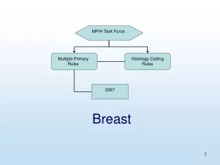
Breast
Breast. Differential diagnosis for breast lump. Malignant lump Breast abscess Fibrocystic changes : Lumpiness, thickening and swelling , often associated with a woman’s period Fibroadenomas : A solid, round, rubbery lump that moves under skin when touched, occuring most in young women


Breast
E N D
Presentation Transcript
Differential diagnosis for breast lump • Malignant lump • Breast abscess • Fibrocystic changes: Lumpiness, thickening and swelling, often associated with a woman’s period • Fibroadenomas: A solid, round, rubbery lump that moves under skin when touched, occuring most in young women • Infections: The breast will likely be red, warm, tender and lumpy • Trauma: a blow to the breast or a bruise can cause a lump (traumatic fat necrosis)
Fibrocystic breast disease(FBD) • Definition: - It is noncancerous breast lumps which can sometimes cause discomfort often periodically related to hormonal influences from the menstrual cycle. • Synonyms: • Cyclic Mastopathy • Cystic Hyperplasia
epidymology • Incidence: • 50% in premenopausal years • FBD represents a clinical problem in approximately 30% of patients.
causes • related periodically to ovarian hormone levels specially if there is predominence of estrogen over progesterone as in anovulation and no enough progesterone production . • estrogen stimulate proliferation of connective and epithelial tissues. • But the condition usually subsides after menopause as estrogen level decrease. • The incidence of FCD is lower in women taking birth control pills having progesterone ,alsois rare in ovulating women, multiparous women.
Diagnosis of FBD • 1- symptoms& signs • 2- investigations • 3-biopsy for histopathology
Diagnosis • 1-Symptoms and Signs • Fibrocystic disease has a history of many months to several years. 1- Breast pain (mastodynia) and/or tenderness is observed in the majority of patients. Mastodynia may start few days before menstruation; it usually subsides during menses. 2- nipple discharge
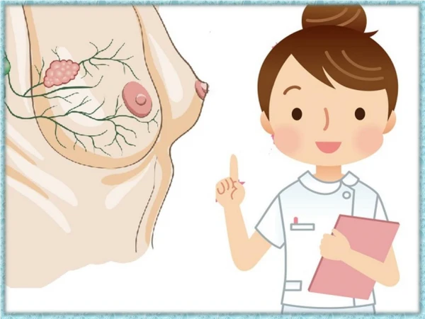
Signs of FBD The lumps are most often found in the upper outer quadrant of the breast (nearest to the armpit). *characterised by the appearance of fibrous tissue and a lump. These lumps are smooth with defined edges, and are usually free-moving in regard to adjacent structures
Histopathology for Fibrocystic Changes Proliferative Change Nonproliferative Change
Histopathology • Oestrogen stimulate proliferation of connective and epithelial tissues.' A- proliferative changes: - lobular hyperplasia: increase number of breast lobules. - fibrosis • cyst formation, • Epithelial proliferation,in form of intraductal epithelial proliferation(papillomatosis). B-regressive changes to the proliferated intralobular connective tissue may undergo : Loss of parenchymal elements (ductules, alveoli) with intra-lobular and periductal fibrosis is encountered in chronic disease.
Pathogenesis of cyst formation • Cyst formation as a consequence of obstruction by stromal fibrosis and persisting ductular alveolar secretion, whereby material is retained, leading to dilation of terminal ducts (duct ectasia) and alveoli with cyst formation. • In 20% to 40% of patients with fibrocystic disease, gross cyst formation is observed. • Macrocysts (>1 cm in diameter) represent an advanced form of fibrocystic disease. They develop in women mainly in their forties and, depending on the degree of fluid filling . • Some of the larger cysts in fibrocystic disease may have a bluish appearance from outside (blue-domed cysts).
Fibrocystic disease of breast: show proliferative changes in form of • -lobular hyperplasia • -fibrosis • cyst formation, • Epithelial proliferation,in form of intraductal epithelial proliferation(papillomatosis • The cyst lining is flattened or absent in some cases. In the center of this image, cysts are lined by apocrine epithelium. Note the focus of adenosis above it
lobular hyperplasia, adenosisi.e increase number of glands (breast acini) .
FBD: show epithelial hyperplasia inside ductules in form of papillomatosis
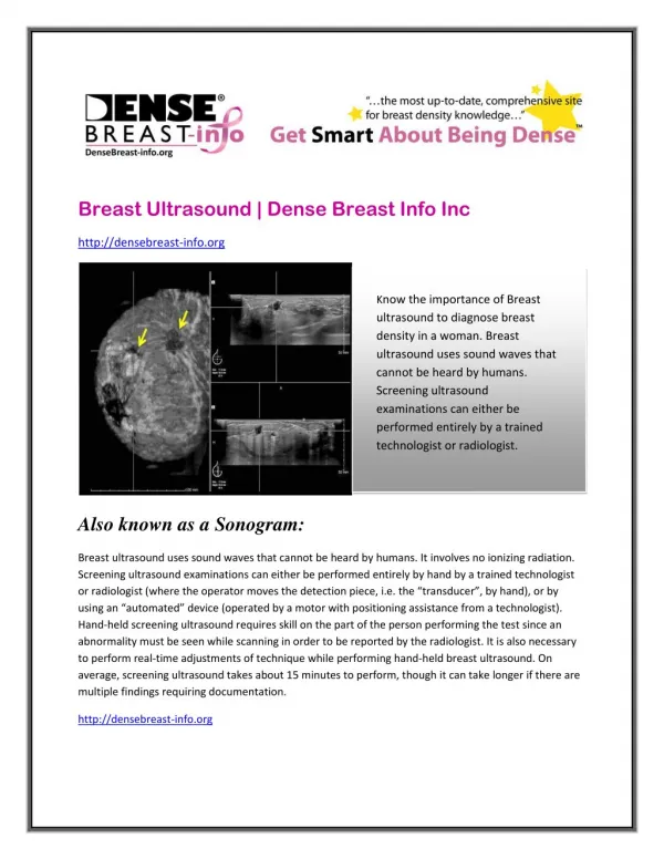
2-investigations 1-Mammography 2- Ultrasonography (USG):Particularly useful in delineating solid from cystic breast masses. • Ultrasound of cystic masses characteristically defines a mass with a uniform outer margin. 3- Needle aspiration biopsy: Indicated in patients with breast mass, a lump like structure,, a hard dense area or any abnormal tissue areas, as defined by clinical examination, mammography or USG. 4- Immunostain for Growth cystic disease fluid protein (GCDFP-15)is strongly positive in FBD.GCDFP is a glycoprotein which is localized in the apocrine metaplastic epithelium lining of breast cysts .
Apocrine metaplasia is a frequent finding in fibrocystic disease. The lining cells have abundant eosinophilic granular cytoplasm, prominent nucleolus and apocrine snouts.
Relation between FBD and cancer breast: A woman who has fibrocystic disease isn't necessarily at risk for developing breast cancer, unless atypical hyperplasia (abnormal cells lining the breast lobules and ducts) is present