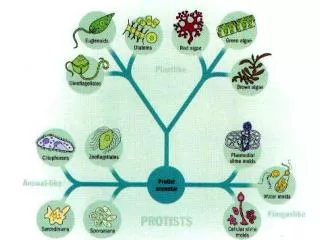
Protozoa
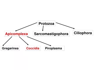
Protozoa. Ciliophora. Apicomplexa. Sarcomastigophora. Gregarinea. Coccidia. Piroplasma. Coccidia. characterized by thick-walled oocysts excreted in feces. In Humans Cryptosporidium Isospora Cyclospora Sarcocystis Toxoplasma. Eimeria. Cryptosporidium. Coccidians:.

Protozoa
E N D
Presentation Transcript
Protozoa Ciliophora Apicomplexa Sarcomastigophora Gregarinea Coccidia Piroplasma
Coccidia • characterized by thick-walled oocysts excreted in feces • In Humans • Cryptosporidium • Isospora • Cyclospora • Sarcocystis • Toxoplasma
Eimeria Cryptosporidium Coccidians: • Eimeria tenella: coccidiosis • Cryptosporidium spp: cryptosporidiosis
Rhoptries and Micronemes are secretory: important in invasion of host cells Microtubules: support-these disappear after parasite is established in the host cell.
Eimeria and Isospora Intestinal The life cycles are similar Infections may be asymptomatic or very pathogenic Coccidia are microscopic parasites detectable on routine fecal tests. Coccidia infection causes a watery diarrhea which is sometimes bloody and can even be a life-threatening problem.
The coccidia have a complex life cycle that includes 3 sequential stages: endogenous merogony and gamogony followed by sporogony which is exogenous. This complexity resulted in various stages of the same coccidian species being described as different species, or even placed in different higher taxa (genera to suborders), before their basic life history was understood. The endogenous (intracellular) developmental stages in a coccidian life cycle are unknown in many / most described species and may be impossible to find or identify under field conditions, so these characters have little present taxonomic value. The exogenous stage (oocyst), upon which the majority of all species descriptions are based, is highly resistant to many fixative techniques and, to date, no satisfactory method is known to permanently preserve all structural features. As a result, most species are described solely on measurements of different structures in the sporulated oocyst, some additional key qualitative features, and line drawings.
Eimeriidae are homoxenous (direct life cycle), with merogony, gamogony and the formation of oocysts occurring within the same host. Oocysts leave the host via the feces, and are unsporulated (undeveloped, non-infective). The development of a genetically-determined number of sporocysts and sporozoites within each oocyst usually occurs outside the host if/when environmental conditions (oxygen, moisture, temperature) are appropriate. The genus Eimeria with more than 1700 species described to date, is the largest apicomplexan genus. Sporulated oocysts of Eimeria contain four sporocysts, each with two sporozoites. Structure and Life History Since the oocyst is the stage that leaves the host, usually in the feces, it is the structure in the life cycle that readily is available to the veterinarian, wildlife biologist, or parasitologist who wants to identify the species, often without having to kill the host. As a result, about 98% of all Eimeria species are known only from this one life-cycle stage, the sporulated oocyst.
Eimeria When a sporulated oocyst is ingested by the proper host, the sporozoites must first leave the confines of the sporocyst and oocyst (they must "excyst") before infection can proceed. Both mechanical (muscular contractions) and enzymatic digestive (trypsin, bile salts) processes of the upper gastrointestinal tract of the host make the sporocyst and oocyst walls more permeable; eventually, certain parts of each may be digested, or they may collapse or are broken, releasing their sporozoites Once free, a sporozoite must penetrate a host epithelial cell before development can continue. Invasion of the host cell involves a sequential series of steps including recognition of a host cell, attachment to surface components, formation of a tight junction, entry into the cell (facilitated by components of the apical complex), and formation of a parasitophorous vacuole around the sporozoite. Safely inside its parasitophorous vacuole, the sporozoite initiates merogony (asexual multiple fission).
Eimeria During merogony 2- 100,000 merozoites may be formed by each sporozoite, depending on the species. Mature merozoites rupture and kill the host cell, each seeking to penetrate a new epithelial cell to begin merogony again. It is this stage of the infection that can result in problems. Pathology is related to intestinal cell destruction. Fever is not common unless secondary infection occurs. Bloody diarrhea is common in some infections with a resulting anemia. In general, the disease caused by coccidia varies with the state of the host, the species of coccidia, the degree of parasitism, and the site of infection. In severely infected animals, death may occur and in others, central nervous system disorders may be notable. It is believed that each Eimeria species is programmed genetically for a specific number of merogonous generations characteristic of that species. For the few species in which the actual number of generations is known, it varies from two to four generations. Thus, infections with Eimeria species are self-limiting as asexual reproduction does not continue indefinitely. Nonetheless, whatever the number, tremendous biological magnification of the parasite results from these developmental stages.
Eimeria When the last generation of merozoites enter host epithelial cells, they develop not into additional meronts, but gamonts: macrogametocytes and microgametocytes, each of which will undergo multiple fission to produce thousands of motile, biflagellated micro- or macro gametes. Micro gametes seek our macrogametes and fertilization occurs, restoring the diploid (2N) condition. Soon after fertilization, a delicate membrane forms around the zygote and two types of wall-forming bodies develop in the cytoplasm; these migrate toward, and then fuse with, the surface membranes to form the resistant oocyst wall. When the oocyst wall is fully formed, the oocyst ruptures from the host cell and leaves the host in the feces. The mechanisms that regulate if a merozoite will become a macro- or microgamont, how microgametes find cells with developed macrogametes, and details of the fertilization process are not known.
Once outside the host, the oocyst must sporulate before it is infective to another host animal. The presence of oxygen, moisture, shade, generally, a temperature less than body temperature of the host, are necessary for oocyst survival. If these conditions are met, division, development, and maturation continue. Moisture, temperature, and direct exposure to sunlight all influence the ability of oocysts to sporulate in the external environment. In general, oocysts sporulate more rapidly at higher temperatures but exposure to temperatures less than 10 º C or greater than 50 º C are lethal to unsporulated oocysts. Once sporulated, oocysts of some species remain viable and infective in 2% aqueous potassium dichromate (kills bacteria, prevents putrefication) at 4-5 º C for up to four years. In their natural external environment, oocysts remain viable and infective from as little as 49 days up to 86 weeks, dependent upon the species and the interplay of abiotic and biotic environmental parameters.
Eimeria Eimeria species demonstrate both site and host specificity, but to somewhat different degrees. The majority of species undergo development within certain cells of the gastrointestinal tract, but not all species are found in this location. Other species have been found to develop in cells of the gall bladder, placenta, epididymis, uterus, genitalia of both sexes, bile duct, liver parenchyma, etc. Once within their specific organ system of choice, Eimeria species seem to be limited to specific zones within that system, specific cells within that zone, and specific locations within those cells. Thus, one species may be found only in the middle third of the small intestine and another only in the cells of the cecum. Within their specific region, one species may be found only in cells at the base of the Crypts of Lieberkühn, a second species in epithelial cell along the villi, and a third species in endothelial cells of the lacteals in the villi.
A histological section showing the asexual reproductive stages of a coccidian in the tissues of the host's small intestine. Note the many developing meronts (=schizonts) (the large dark blue structures enclosed within the rectangle) in the tissues. Each meront will produce many merozoites
This is a typical life cycle of intestinal coccidia in birds
Bloody diarrhea, sloughing of epithelium, cell death, affect uptake, cause hemorrhage Sub lethal effects may predispose wild birds to predation or render commercial operations non functional
(A) Hemorrhage in the small intestine of a lesser scaup with acute intestinal coccidiosis (upper part of photo), compared with normal small intestine (lower part of photo). (B) Dry, crust-like lesions in the intestinal tract of a lesser scaup with chronic intestinal coccidiosis. The lesions are most severe in the upper small intestine (top section in photo). The severity decreases in lower parts of the intestine (middle and bottom sections in photo).
Cryptosporidium parvum • First human case reported in 1976 • Became important emerging disease in humans in 1980s in immunocompromised people • Lumped in with the other coccidia but they are different. • monoxenous, wide range of animal hosts • several host-adapted species? • self-limiting diarrhea in immunocompetent hosts • profuse, watery diarrhea associated with AIDS- life threatening • associated with epidemic diarrhea in institutions and hospitals • highly transmissible • Several outbreaks: seroprevalence is often high indicating exposure: blood donors in Edinburgh 86% prevalence
Cryptosporidium Life Cycle • Type I meronts produce 6-8 merozoites, invade, become Type II • Type II merozoites produce 4 merozoites, many of which enter gametogony • Fertilization- oocysts- sporulation occurs within tissues • Oocysts can exit with feces or excyst in intestine and begin all over again
Extracytoplasmic Location • microvilli extend and fuse to enclose zoite • close association between parasite and host intestinal epithelial cell • called adhesive zone, feeder organelle, etc
Pathogenesis • DIARRHEA • enterocyte malfunction (osmotic) • impaired absorption • enhanced secretion • inflammatory diarrhea • mucosal invasion • leukocytes in stools • secretory diarrhea • toxin • watery • enterocytes damaged or killed • villus atrophy (blunting) • Na+ absorption • permeability • crypt cell hyperplasia • Cl- secretion • inflammation
The Milwaukee Outbreak NEJM 331:161 (1994) • massive cryptosporidiosis outbreak following spring thaw • >400,000 people may have been affected • based on clinical symptoms (acute watery diarrhea) • treated water had high levels of turbidity 3/23-4/5/1993 • oocysts identified in ice made during this period • 100-fold higher prevalence of Cryptosporidium oocysts in stools • other enterics (including Giardia, bacteria, viruses) were at ~normal levels
Factors Favoring Waterborne Cryptosporidiosis • small size of oocysts (4-5 mm) • reduced host specificity and monoxenous development • close associations between human and animal hosts • large number of oocysts excreted (up to 100 billion per calf per day) • low infective dose (<30) • robust oocysts; resistant to chlorine • infectious sporulated oocysts excreted in feces
Two distinct transmission cycles: • anthroponotic (genotype 1) • zoonotic (genotype 2)
Intestinal Coccidia • Diagnosis • demonstration of oocysts in feces • acid-fast stain • autofluorescence (Isospora and Cyclospora) • direct observation (Isospora) • Treatment • paromomycin for Cryptosporidium • modest benefit • lowers parasitemia in AIDS • trimethoprim-sulfamethoxazole for Cyclospora and Isospora