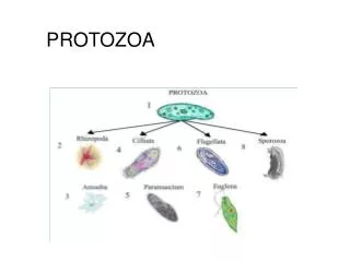
Protozoa
Protozoa. Protozoa, the tree of life and the origin of eukaryotes What makes an ameba an ameba? Entamoeba histolytica infection and pathogenesis. protozoa. Primary unicellular eukaryotes, often also called protists Many important human and veterinary pathogens

Protozoa
E N D
Presentation Transcript
Protozoa • Protozoa, the tree of life and the origin of eukaryotes • What makes an ameba an ameba? • Entamoeba histolytica infection and pathogenesis
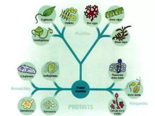
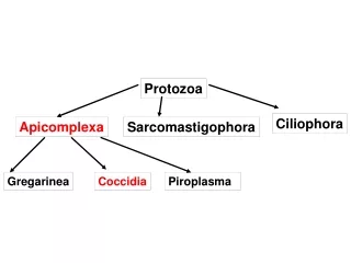
protozoa • Primary unicellular eukaryotes, often also called protists • Many important human and veterinary pathogens • A very diverse group with a vast variety of morphological and biochemical adaptations to almost any ecological niche • Where do they come from, how do they relate to each other, and how do they relate to us?
From taxonomy to the tree of life (Linneus to Haeckel) • Taxonomy classifies organisms into meaningful groups that help to conquer and understand the massive diversity • The tree of life concept uses evolution as guiding principle of taxonomy • No evolution – no tree. • Choosing the tree model makes several important assumptions • All life is related • Life diversifies • Life has a common origin
the tree of life(Ernst Haeckel, 1874) man ungulates • The tree of life (who is related and how did they evolve) was initially based on anatomical characteristics • “Complex” organisms were viewed as derived and highly evolved “simple” organisms (like the ameba) as primitive • This scheme puts protozoa to the bottom of the tree and animals to the top carnivores whales fish reptiles crustaceans molluscs worms protozoa
the tree of life(Ernst Haeckel, 1866) • Monophyletic tree of organisms again by Haeckel • Loss or gain of characters produces branching of the tree • The advent of electron microscopy brought more morphological characters even for the small protists • However, reduction and simplifications (e.g. due to parasitism) pose significant problems for morphology based trees • Furthermore homology is not always discernable from analogy, and characters are not always easily quantifiable
Molecular phylogeny • Uses the sequence of macromolecules (RNA, DNA & proteins) to measure similarity, and to deduce evolutionary relation • Informative molecules are present in all the organisms to be compared • Relatedness is inferred from the simple argument that two molecules from two related organisms are likely to be more similar than from two organisms that diverged a long time ago 30S ribosomal subunit, rRNA pink Schluenzen et al. Cell 102 (5): 615–23.
Molecular phylogeny • Molecular phylogeny assumes that sequence changes occur over time and that these changes can be modeled and used to infer a process (evolution) out of the current pattern
Molecular phylogeny • A large number of statistical approaches has been developed to compare sequences, build trees that depict the results, and evaluate their statistical significance • A tree of life based on the ribosomal RNA sequence • Three major domains (two prokaryotic one eukaryotic)
The archezoa hypothesis • Eukaryotes have some features of bacteria, some of archaea and some new ones • How did they evolve? • A key event was the acquisition of the mitochondrion (we will discuss endosymbiosis in detail in a later class) but when did that occur? • Who is the “most primitive eukaryote”
Archezoa & amoeba the most primitive eukaryotes? • No mitochondrion and no typical mitochondrial enzymes (Krebs cycle and oxidative phosphorylation is missing) • It was assumed that archezoa and amoeba represent the stage of early eukaryotes before the endosymbiosis event that let to the mitochondrion • An alternative hypothesis stated that these organisms once had mitochondria and subsequently lost them while adapting to parasitism and life in anaerobic environments
Is the absence of mitochondria a primary of secondary trait? • The genomes of most important protozoan parasites are now fully sequenced • This provides the opportunity to hunt for ‘molecular fossils’ • Most proteins that do their job in the mitochondrion are actually encoded in the nucleus and are imported from the cytoplasm • Genome searches in Entamoeba and Giardia identified several genes for proteins targeted to the mitochondrion in other organisms
Is the absence of mitochondria a primary of secondary trait? • Localizing these proteins identified a small highly reduced mitochondrion - the mitosom • This suggest amoebae had full-fledged mitochondria in the past but reduced them when they moved to an anaerobic environment • For the moment we don’t know a eukaryote featuring a primary lack of mitochondria Cpn60 DIC Microbiology 150 (2004), 1245-1250
Amoeboid movement Acanthamoeba http://cmgm.stanford.edu/theriot/movies.htm#Hits
what is amoeboid about amoebae? Pseudopodium Hyaline ectoplasm (gel) Endoplasm (sol) While the endoplasm is ‘liquid’ and filled with organelles the ectoplasm appears ‘solid’ and clear. Uroid
Amoeboid movement is not limited to amoeba Neutrophil chasing a bacterium What could be the engine and gears powering this movement?
Ameboid movement is driven by actin • Amoeboid movement depends on the actin cytoskelleton • Earlier models were based on cortical actin/myosin squeezing the cytoplasm to the leading edge (toothpaste tube model), this was thought to be accompanied by cytoplasmic gel/sol transformations • More recent data strongly support actin polymerization as the force generating step (at least for the best understood part of protrusion) • Actin dynamics in amoeboid movement are complex and not easily dissected -- can just polymerizing actin really drive motility?
Listeria as a model to demonstrate and study actin polymerization motility • The actin polymerization model is based on cell free reconstitution of the movement of intracellular bacteria • These studies allowed to identify the factors involved in the initiation of actin filament polymerization Listeria in Xenopus extract (right panel Phase contrast, left panel actin-GFP fluorescence) Listeria in host cell (150x) http://cmgm.stanford.edu/theriot/movies.htm#Hits
Entamoeba histolytica • Fedor Alexandrewitch Lösch describes amoebae associated with severe dysentery in a patient in 1873 • He transferred amoebae to a dog by rectal injection, which became ill and showed ulceration of colon • Patient who died from infection showed similar ulcers upon autopsy
trophozoites and cysts • multiple well defined pseudopodia often extended eruptively • Differentiation into endo- and ectoplasm • Spherical nucleus (4-7 mm) with small central nucleolus and characteristic radial spokes
trophozoites and cysts • Trophozoites 20-40 mm diameter • Ribosomes arranged in helical patterns • Tissue forms often contain phagocytized red blood cell
trophozoites and cysts • Trophozoites encyst and cysts mature as they travel through the colon • Only mature cysts are infective
trophozoites and cysts • Round (10- 16 mm), 4 nuclei • 150 nm cyst wall with fibrillar structure • Chromidial bodies and bars are semicrystalline arrays of riobosomes
Entamoeba cysts (light microscopy) E. coli E. histolytica
Human infection • Major sources for human infection are contamination of drinking water and vegetables (fertilization with material containing or contaminated with human feces) • Patients without any symptoms might nevertheless shed large amounts of cysts • If kept cool and moist (water or soil) cysts can stay infectious for up to a month • Cysts are fairly resistant to chlorination of drinking water (10 mg/l versus 0.1 - 1.0 mg/l for enteric bacteria)
Colitis is the most common form of disease associated with amoebae • Gradual onset of abdominal pain, watery stools containing mucus and blood • Some patients have only intermittent diarrhea alternating with constipation • Fever is uncommon • Formation of ulcers
Colitis is the most common form of disease associated with amoebae • Amoeba invade mucosa and erode through laminia propria causing characterisitic flask shaped ulcers contained by muscularis
Ulceration can lead to secondary infection and extraintestinal lesions
Amebic liver abscess • Most common form of extraintestinal amebiasis • Fast growing abscess filled with debris, amoebae are found only at borders • Lead symptoms are are right upper quadrant pain and fever • Acute as well as chronic illness, with gradual or sudden onset
Amebic liver abscess • 30-50% of patients with liver abscess show also pneumonic involvement • Rupture is again a major thread, especially rupture into the pericardium • Draining abscesses is today only performed in extreme cases when rupture is feared • Responds well to chemotherapy
Metronidazole is the drug of choice for extra-intestinal amebiasis • Several drugs are available to clear symptomatic and asymptomatic enteric (luminal) infection (e.g. dichloroacetamides which have unknown mode of action) • Metronidazole (Flagyl) is the drug of choice for invasive amoebiasis (and should be combined with a lumen acting drug as it is not fully effective on luminal stages) • Metronidazole is a prodrug which is activated by an enzyme involved in the fermentative metabolism of E. histolytica (PFOR – Dr. Moreno will explain this in the next lecture)
Amoebae use fermentation • “La fermentation est la vie sans l’air” (Louis Pasteur) • Entamoeba lacks a functional Krebs cycle and oxidative phosphorylation • Final endproducts of E. histolytica fermentation are CO2, acetate, ethanol and alanine
Metronidazole is activated by PFOR • Entamoeba uses a pyruvate ferredoxin oxidoreductase (PFOR) to break down pyruvate • This process depends on the absence (or low level) of oxygen • This enzyme system is limited to anaerobic bacteria and some protozoa and humans lack this enzyme • PFOR and ferredoxin can transfer an electron to metronidazole producing a highly toxic nitroradical • Drugs which are not toxic but have to be activated into a toxic compound are called prodrugs (look at this as a suicide substrate)
Epidemiology of Entamoeba • 480,000,000 people harbor Entamoeba • 36,000,000 develop clinical symptoms • 40,000 - 100,000 deaths per year (Walsh, 1986, Rev. Infect. Dis., based on 1981 data, no significant change since then) Less than 10% of the people infected show disease. Several hypotheses have been put forward to explain this differential pathogenesis.
Commensal hypothesis • E. histolytica usually is a benign gut commensal as many other amoebae (minuta form) • A certain stimulus (gut flora, diet, host immune status …) transforms the organism into a pathogen (magna form, Kuenen, 1913) • This has been the accepted view for most of the 20th century
Two species hypothesis • There are two morphologically indistinguishable species: E. histolytica and E. dispar. Only one of them causes (Brumpt, 1928) • This theory was discounted • Recent molecular data have revived the two species hypothesis • We now know that most people are infected with the apathogenic E. dispar • How do ameba cause disease? Emile Brumpt 1877-1951
Entamoeba pathogenesis factors • What are pathogen proteins (and genes) that are required to cause disease? • Several candidates have been studied for their involvement in contact dependent cell killing by amoeba: • The surface lectins: These are proteins that allow the amoeba to bind to sugars on the surface of cells and establish tight contact • Proteases: several protein degrading enzymes have been linked to tissue penetration and liver abscess formation • Amoebapores: protein toxins that perforate target cells
Amoebapores one of the candidate pathogenicity factors • Family of small (77 AA) proteins contained in secretory granules • Similar in structure and function to NK lysins • Used to kill bacteria and host cells • Amoebapores insert into target membranes and form ion channels • Amoeba mutants which make less amoebapores cause less disease in animal model studies