PET Phantom Segmentation Challenge: Part 2 - Analysis and Measurement Instructions
80 likes | 220 Vues
In Part 2 of the PET Phantom Segmentation Challenge, participants will perform segmentations and analyses on the UI and UW PET phantom volumes. Known volumes and activities are provided for accuracy. Segmentations must be repeated twice for all datasets using the Iowa PET phantom data from Dr. Sunderland. Participants are required to adhere to a specific naming convention for reporting measurement results. Analysis includes generating volumetric segmentations, calculating various indices, and identifying the actual boundaries of the objects in the scans.

PET Phantom Segmentation Challenge: Part 2 - Analysis and Measurement Instructions
E N D
Presentation Transcript
PART 2: QIN PET Phantom Segmentation Challenge Instructions
Goals for Part 2 • Perform segmentations and analysis on the UIand UW PET phantom volumes • Volumes and activities are known • Two repeat segmentations/measurements for all data sets!
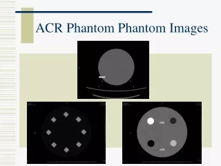
Iowa PET phantom data from Dr. Sunderland: Part 2: PET Phantom Image Data - NEMA IEC Body Phantom Set™ Model PET/IEC-BODY/P with unknown sized spheres & ellipses. - Downloadable from website.
Iowa PET Phantom Data Four Image Sets per Site
Directory Structure and File Names Site/Scanner PET Iowa (Siemens Scanner) Scan Type PET Washington (GE Scanner) 138M ./UW 13M ./UW/HS 6.3M ./UW/HS/LC 6.3M ./UW/HS/HC 125M ./UW/LS 63M ./UW/LS/LC 6.3M ./UW/LS/LC/REBIN_A_4I28S3MM_406 6.3M ./UW/LS/LC/REBIN_B_4I28S3MM_407 6.3M ./UW/LS/LC/REBIN_C_4I28S3MM_408 6.3M ./UW/LS/LC/REBIN_D_4I28S3MM_409 6.3M ./UW/LS/LC/REBIN_E_4I28S3MM_410 6.3M ./UW/LS/LC/REBIN_F_4I28S3MM_411 6.3M ./UW/LS/LC/REBIN_G_4I28S3MM_412 6.3M ./UW/LS/LC/REBIN_H_4I28S3MM_413 6.3M ./UW/LS/LC/REBIN_I_4I28S3MM_414 6.3M ./UW/LS/LC/REBIN_J_4I28S3MM_415 63M ./UW/LS/HC 6.3M ./UW/LS/HC/REBIN_A_4I28S3MM_406 6.3M ./UW/LS/HC/REBIN_B_4I28S3MM_410 6.3M ./UW/LS/HC/REBIN_C_4I28S3MM_412 6.3M ./UW/LS/HC/REBIN_D_4I28S3MM_415 6.3M ./UW/LS/HC/REBIN_E_4I28S3MM_418 6.3M ./UW/LS/HC/REBIN_F_4I28S3MM_421 6.3M ./UW/LS/HC/REBIN_G_4I28S3MM_424 6.3M ./UW/LS/HC/REBIN_H_4I28S3MM_427 6.3M ./UW/LS/HC/REBIN_I_4I28S3MM_430 6.3M ./UW/LS/HC/REBIN_J_4I28S3MM_433 105M ./UI 9.6M ./UI/HS 4.8M ./UI/HS/LC 4.8M ./UI/HS/HC 96M ./UI/LS 48M ./UI/LS/LC 4.8M ./UI/LS/LC/Frame01_LC 4.8M ./UI/LS/LC/Frame02_LC 4.8M ./UI/LS/LC/Frame03_LC 4.8M ./UI/LS/LC/Frame04_LC 4.8M ./UI/LS/LC/Frame05_LC 4.8M ./UI/LS/LC/Frame06_LC 4.8M ./UI/LS/LC/Frame07_LC 4.8M ./UI/LS/LC/Frame08_LC 4.8M ./UI/LS/LC/Frame09_LC 4.8M ./UI/LS/LC/Frame10_LC 48M ./UI/LS/HC 4.8M ./UI/LS/HC/Frame01_HC 4.8M ./UI/LS/HC/Frame02_HC 4.8M ./UI/LS/HC/Frame03_HC 4.8M ./UI/LS/HC/Frame04_HC 4.8M ./UI/LS/HC/Frame05_HC 4.8M ./UI/LS/HC/Frame06_HC 4.8M ./UI/LS/HC/Frame07_HC 4.8M ./UI/LS/HC/Frame08_HC 4.8M ./UI/LS/HC/Frame09_HC 4.8M ./UI/LS/HC/Frame10_HC high statistics, low contrast high statistics, high contrast 10 x low statistics, low contrast 10 x low statistics, high contrast
Object Naming Convention • For the reporting of measurement results, etc. participants must adhere to the following naming convention! 6 6 5 5 start 4 4 1 1 2 2 3 3 clock wise flat part of phantom Axial PET slice Volume Rendering
PET Phantom Data • Perform the following analysis steps for objects 1 to 6: • Generate accurate volumetric segmentations of the objects in the phantom scans • Segment the VOIs in such a way as to identify the actual boundaries label VOIs (e.g., VOI1, VOI2, ...) • Calculate the following indices for each of the objects: • VOI volume in [ml] • MAXIMUM Concentration in [Bq/mL] • PEAK Concentration (PERCIST; 1 cm sphere???) in [Bq/mL] • AVERAGE Concentration in [Bq/mL] • Metabolic Tumor Volume (Average Concentration * VOI Volume) in [Bq]


![[PDF⚡READ❤ONLINE] Phantom](https://cdn7.slideserve.com/13158923/slide1-dt.jpg)