The Cranial Nerves XI-XII
271 likes | 1.15k Vues
The Cranial Nerves XI-XII. Dr. Zeenat Zaidi Dr.Essam Eldin Salama. Objectives . At the end of the lecture, the students should be able to: List the nuclei related to accessory and hypoglossal nerves in the brain stem. Describe the type and site of each nucleus.

The Cranial Nerves XI-XII
E N D
Presentation Transcript
The Cranial NervesXI-XII Dr. ZeenatZaidi Dr.EssamEldinSalama
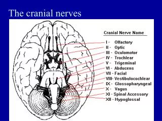
Objectives At the end of the lecture, the students should be able to: List the nuclei related to accessory and hypoglossal nerves in the brain stem. Describe the type and site of each nucleus. Describe site of emergence and course of accessory and hypoglossal nerves. Describe important relations of accessory and hypoglossal nerves in the neck. List the branches of accessory and hypoglossal nerves. Describe the main motor effect in case of lesion of accessory and hypoglossal nerves.
11th CN: Accessory Nerve Type: Motor Has two parts: Cranial Spinal Foramen of exit from skull: Jugular foramen
Cranial Part Origin:Nucleus ambiguus in the medulla oblongata. Type of fibers: SVE Site of emergence: The fibers emerge from the anterior surface of the medulla oblongata between the olive and the inferior cerebellar peduncle (ICP).
Spinal Part Origin: It is formed by the axons of the nerve cells in the spinal nucleus. It carries SVE fibers, (embryology) It is located in the ventral grey horn in the upper 5 cervical segments.
Accessory Nerve The nucleus ambiguus and the spinal nucleus receive bilateral corticonuclear fibers (afferent) from both cerebral hemispheres.
Accessory Nerve Course: Cranial part: runs laterally and joins the spinal part. Spinal part: fibers emerge from the spinal cord, form a nerve trunk that ascends into the skull through the foramen magnum, passes laterally and joins the cranial root.
Accessory Nerve The two roots unite and exit through the jugular foramen. The cranial part separates from the spinal part and joins the vagus nerve. The spinal part runs downwards to supply muscles of neck (sternomastoid & trapezius)
Accessory Nerve Distribution: Cranial part: distributed through vagus nerve, to muscles of larynx, pharynx, soft palate & esophagus Spinal part: enters deep surface of sterno-cleidomastoid muscle, supplies this muscle and then crosses the posterior triangle of the neck and supplies the trapezius muscle.
Accessory Nerve Function: Movements of the soft palate, larynx, pharynx. Controls the movements of neck Lesion results into: Difficulty in swallowing and speech Inability to turn the head and raise the shoulder Winging of scapula
12th CN: Hypoglossal Nerve XII nucleus Type: Motor Origin: Hhypoglossal nucleus of the medulla (in the floor of 4th ventricle) Type of fibers: GSE. Fibers emerge between pyramid and olive from ventral aspect of medulla. olive pyramid
Hypoglossal Nerve Site of emergence: The fibers emerge from the anterior surface of the medulla oblongata between the pyramid and the olive. Foramen of exit from skull: Hypoglossal canal
Hypoglossal Nerve • The hypoglossalnucleus receives; • Corticonuclear fibers (afferent) from both cerebral hemispheres EXCEPT the region that supplies genioglossus muscle (it receives contralateral supply only). • Afferent fibers from nucleus solitarius and trigeminal sensory nucleus.
Hypoglossal Nerve Course: Descends downward with cervical neurovascular bundle (internal carotid artery, internal Jugular vein, vagus nerve) Curves forward behind mandible to supply the tongue. During its initial course, it carries C1 fibers which leave in a branch to take part in the formation of ansa cervicalis.
Hypoglossal Nerve Distribution: Supplies motor innervations to all of the muscles of the tongue except the palatoglossus (which is supplied by the vagus). Carries proprioceptive afferents from the tongue muscles.
Hypoglossal Nerve Function: Controls the movements and shape of the tongue during speech and swallowing. Lesion; (LMN paralysis) results into: Loss of tongue movements Difficulty in chewing and speech The paralyzed tongue, atrophies, becomes shrunken and furrowed on the affected side. Unilateral lesion; the protruded tongue deviates to the affected side. Bilateral lesion; the person can’t protrude the tongue.