Download
1 / 67
710 likes | 1.88k Vues
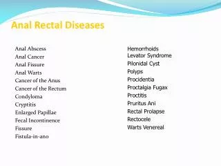
Anal Rectal Diseases. Hemorrhoids Levator Syndrome Pilonidal Cyst Polyps Procidentia Proctalgia Fugax Proctitis Pruritus Ani Rectal Prolapse Rectocele Warts Venereal. Anal Abscess Anal Cancer Anal Fissure Anal Warts Cancer of the Anus Cancer of the Rectum Condyloma Cryptitis

E N D
Anal Rectal Diseases HemorrhoidsLevator Syndrome Pilonidal Cyst Polyps Procidentia Proctalgia Fugax Proctitis Pruritus Ani Rectal Prolapse Rectocele Warts Venereal Anal Abscess Anal Cancer Anal Fissure Anal Warts Cancer of the Anus Cancer of the Rectum Condyloma Cryptitis Enlarged Papillae Fecal Incontinence Fissure Fistula-in-ano
Anorectal Anatomy Nerve Supply Sympathetic: Superior hypogastric plexus Parasympathetic: S234 (nerviergentis Pudendal Nerve: Motor and sensory Arterial Supply Inferior rectal A middle rectal A Venous drainage Inferior rectal V middle rectal V 3 hemorrhoidal complexes L lateral R antero-lateral R posterolateral Anal canal Lymphatic drainage Above dentate: Inf. Mesenteric Below dentate: internal iliac Anal verge
External • Pain? • -> painless • Bright red bleeding • Prolapse associated with defecation Internal • Anoderm • Swell, discomfort, difficult hygiene • Pain? • -> Thrombosed
HaemorrhoidsBack Ground • They are part of the normal anoderm cushions • They are areas of vascular anastamosis in a supporting stroma of subepithelial smooth muscles. • The contribute 15-20% of the normal resting pressure and feed vital sensory information . • 3 main cushions are found • L lateral • R anterior • R posterior • But can be found anywhere in anus • Prevalence is 4% • Miss labelling by referring physicians and patients is common This combination is only in 19%
HaemorrhoidsPathogensis 3 main processes: 1. Increased venous pressure 2. Weakness in supporting fibromuscular stroma 3. Increased internal sphincter tone Risk Factors
A:Thrombosed external B:First-degree internal viewed through anoscope C:Second-degree internal prolapsed, reduced spontaneously D:Third-degree internal prolapsed, requiring manual reduction E:Fourth-degree strangulated internal and thrombosed external Reference : Sabiston Textbook of Surgery, 18th Edition
HaemorrhoidsInvestigations: • Lab: CBC / Clotting profile/ Group and save • Proctography: if rectal prolpse is suspected • Colonoscopy: if higher colonic or sinister pathology is suspected
Complications • Ulceration • Thrombosis • Sepsis and abscess formation • Incontinence Thrombosed internal haemorrhoids Thrombosed external haemorrhoids
HaemorrhoidsExternal H. Treatment : • If presentation less than 72 hours: • Enucleate under LA or GA • Leave wound open to close by secondary intension • Apply pressure dressing for 24 hours post op • If more than 72 hours: • Conservative measures
Perianal Fistula and Abscess 5% 60% 5% Ischiorectal 20% Intersphincteric suprasphincteric extrasphincteric Trans-sphincteric
Peri-anal FistulaClinical presentation Godsalls law Anterior: drain straight Posterior: drain curved to anorectal midline • Follow 40-60% of perianal abscess and cryptgland infections • Presentation: • External openings • Purulent discharge • Blood • Perianal pain
Perianal AbscessManagement Aim: adequate drainage of abscess preservation of sphincter function * Preop: full lab evaluation *Always perform Examination under GA ( EUA) and obtain a biopsy.
Perianal fistulaManagment Aim: Define anatomy Eliminate tract preservation of sphincter function * Preop: full lab evaluation *Always perform Examination under GA ( EUA) and obtain a biopsy.
Anal Fissure • Linear tears in the anal mucosa exposing the internal sphincter • 90% are posterior • Caused mainly by trauma ( hard Stool). Followed by increased sphincter tone and ischemia. • Other causes: IBD, Ca, Chronic infections
Pilonidal Sinus Pathogenesis: A sinus tract at natal cleft resulting from: • Blockage of hair follicle • Folliculitis • Abscess followed by sinus formation. • Hair trapping • Foreign body reaction • The sinus tract is cephald Associated with: • Caucasians • Hirsute • Sedentary occupations • Obese • Poor hygeine
Presentation & Treatment • Also found: umbilicus, finger webs, perianal area
History • Age • Hemorrhoids- • common all ages but are uncommon below the age of 20 years. • Perianal haematomata- • occurs at all ages • Fissure-in-ano-(acute) • quite common in children • Anorectal abscess- • common between the ages of 20 and 50 years. • Pilonidal sinus- • rare before puberty and in people over 40 years.
History • Sex • Hemorrhoids- • common in both sexs • Perianal haematomata- • occurs at all ages • Fissure-in-ano- • common in men • Anorectal abscess- • more common in men • Pilonidal sinus- • more common in men • Prolapse of rectum- • more common in women
History • Principal symptoms of rectal and anal conditions: • Bleeding • Pain • Tenesmus • Change in bowel habit • Change in the stool • Discharge • pruritis
History - Bleeding • Can be fresh or altered • Example of altered is melaena • Black tarry stool • Recognizable blood may appear in four ways: • Mixed with faeces • On the surface of the faeces • Separate from the faeces: after/unrelated to defaecation • On the toilet paper after cleaning
History - Bleeding • Diagnosis of anal conditions which present with rectal bleeding • Bleeding but no pain: • Blood mixed with stool = ca of colon • Blood streaked on stool = ca of rectum • Blood after defaecation = hemorrhoids • Blood and mucus = colitis • Bleeding + pain = fissure or carcinoma of anal canal • The most common causes of rectal bleeding in patients who visit primary care physicians are hemorrhoids, fissures and polyps.