Download
1 / 54
540 likes | 776 Vues
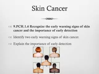
A Clinical Overview of Skin Cancer. Rich Callahan MSPA, PA-C ICM I – Summer 2009 . Recognition and treatment of skin cancer will only become more important for those going into primary care, geriatrics and dermatology. You will see it often in your career.

E N D
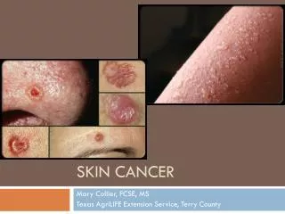
A Clinical Overview of Skin Cancer Rich Callahan MSPA, PA-C ICM I – Summer 2009
Recognition and treatment of skin cancer will only become more important for those going into primary care, geriatrics and dermatology • You will see it often in your career. • By far the most common cancer, and one of the few whose incidence is still increasing dramatically. • Incidence generally increases with age – our aging population structure will ensure a steady supply of cases over time. • Goal is prevention and early recognition. A small skin cancer diagnosed early will generally create much less overall morbidity than a larger skin cancer diagnosed late.
Incidence and Epidemiology(From www.aad.org) • Approximately 1.2 million cases this year. • 80% Basal Cell Carcinoma (BCC) • 16% Squamous Cell Carcinoma (SCC) • 4% Malignant Melanoma (MM) • <1% are the extremely rare skin cancers: Desmoplastic melanoma, eccrine porocarcinoma, cutaneous T-Cell lymphoma (CTCL,) sebaceous gland carcinoma, Kaposi’s sarcoma, leukemia cutis, angiosarcoma, etc. • We will focus on the big 3 because you will see these in clinical practice.
Skin Cancer – The Big 3 • BCC: The most common and least aggressive skin cancer. Slowly growing with low potential to metastasize and/or invade underlying structures. Lesions can be subtle and insidious, infiltrating large areas before they are discovered. Huge range of potential presentations. • SCC: 2nd most common skin cancer. More aggressive than BCC, with higher potential to invade underlying structures if not treated promptly. Usually, it is the most advanced cases that metastasize, but lesions on the lip, nose and ear will do it earlier and must be recognized! Actinic Keratosis (AK) is a precursor lesion to SCC. • BCC/SCC referred to collectively as non-melanoma skin cancer, or NMSC, which accounts for approx. 2,200 deaths/year and huge surgical morbidity.
Breakdown of the big 3 into subtypes • BCC: Nodular, infiltrative, superficial, morpheaform (sclerosing,) and fibroepithelioma of Pinkus. • SCC: Invasive, SCC in situ (Bowen’s Disease.) • MM: MM in situ, superficial spreading, nodular, amelanotic and lentigo maligna.
Epidemiology/Risk Factors for NMSC • People at highest risk: • Fair skin (Fitzpatrick I or II,) and history of excessive, unprotected sun exposure/sun burns. • Family history of one or more 1st degree relatives with skin cancer (a strong predictor.) • Immune compromise: Can see elderly patients suddenly develop crops of skin cancers as their immune function declines. Also seen in organ transplant recipients (OTR’s) who have several risk factors for skin cancer and who are on strong immunosuppressive drugs to prevent organ transplant rejection.
Fitzpatrick’s Skin Phototypes • Type 1: Fair skinned. Always burn (even short exposure time of <30 min.,) never tan. • Type 2: Fair skinned. Often burn, tans with great difficulty. • Type 3: Some sunburn at first, but then tan deeply. • Type 4: Never burns, tans with ease. • Types 5-6: Darker skin types which burn only under the most extreme, prolonged exposures.
Malignant Melanoma (MM) Least common of the big three (approx. 56,000 cases/year) but accounts for large majority of skin cancer mortality (approx. 7,700 deaths/year.) Incidence of MM increasing alarmingly in certain groups: #1 cause of cancer death in women 25-30 y/o #2 cause of cancer death in women 30-35 y/o after Breast Ca Quickly becoming a leading cause of cancer death in men 35-45 y/o One AAD publication stated that incidence of MM has increased 7000% since the 1920’s in the U.S.
MM • High cure rate when caught early, but terrible prognosis if it has already started to spread when diagnosed. • Prognosis based on depth of lesion at time of diagnosis. • Deeper tumor = higher chance for metastasis • Metastatic melanoma has high rate of mortality over a 5 year period after diagnosis. • Many chemotherapeutic and immune-stimulating therapies have been tried in treatment, with poor results so far. • Our best defense is early detection.
Risk Factors for MM • Fair skin (Fitz I/II,) blue eyes, red hair, freckles. • History of severe, intermittent sun exposure rather than gradual (think Vermonter going to St. Croix in January.) • History of severe, blistering sun burns before the age of 18. • Presence of dysplastic nevus syndrome • Family history of MM, especially 1st degree relatives who got it young (i.e., in their 30’s/40’s,) as this suggests a genetic vulnerability. • Prior personal history of MM
Why the big increase in MM? • Demographic trends: Over time we have become a more prosperous, mobile society with more free time to sit in the sun and travel to warm climates. The tan look has become synonymous with vitality, well-being and sex appeal. • Clothing: Since the beginning of the 20th century, our culture has trended towards wearing less clothing in public (100 years ago common modesty dictated that you went swimming almost fully clothed, now we have bikini thongs.) • Tanning Beds: Depending on which study you look at, emit between 2-15 times more UV radiation than the sun!
The Single Greatest Risk Factor for Skin Cancer? • Prior personal history of Skin Cancer. • A patient with a past history has a much greater risk for developing additional skin cancers when compared to the general population. • For this reason, we follow the AAD guidelines for follow-up examination of skin cancer patients: • NMSC patients get f/u skin exams at 6 months and one year. • MM patients get f/u skin exams every 3 months for one year!
Clinical Pearl: When you see skin cancer, suspect more skin cancer. • A patient with a new skin cancer diagnosis must get a full-body skin check within 1-3 months of initial diagnosis, to make sure nothing is getting missed. • People with skin cancer tend to get more skin cancer, sometimes even in cases where the patient practices diligent sun protection after the initial diagnosis. • Routine surveillance is critical in this population. The highest morbidity is seen in skin cancer patients who avoid the dermatologist’s office for years at a time.
Precursors to skin cancer • Actinic Keratosis (AK) Precancerous dysplasia and hyperkeratosis of the epidermis induced by UV radiation. A precursor to SCC. • Usually appears first on Fitz 1 or 2 patients in their 30’s and 40’s. • About 33% of untreated AK’s progress to SCC over a person’s lifetime. • Presents as erythematous to brown-pigmented, gritty, rough macules and patches. Usually on scalp, face and forearms.
Precursors to skin cancer • Dysplastic Nevus Syndrome (DNS) - Dysplastic nevi are clinically atypical moles which share some traits of MM both on gross inspection and histologically: Asymmetry of color/structure on gross inspection, and dysplasia/architectural disorder of melanocytes when viewed under microscope. • DNS patients have increased lifetime risk for MM and should be followed closely.
Clinical Presentation of BCC by subtype. • Usually located on head or upper trunk/extremities. • Nodular BCC – a firm, slightly to somewhat raised papule or nodule having a pearly pink to grayish coloration. Sometimes random splotches of pigment and arborized blood vessels are visible. Often ulcerates/bleeds. • Superficial BCC – a shiny, slightly indurated macule, patch or plaque having pearly pink coloration. • Infiltrative BCC – the hardest to cure completely. Same presentation as nodular BCC except base of tumor radiates multiple roots into surrounding skin. The reason Mohs surgery was invented.
More BCC • Morpheaform, or sclerosing BCC – most subtle, insidious and potentially destructive subtype. Presents as an indurated, smooth to slightly depressed off-white plaque. Can grow for years before being noticed. Often the only clue on physical exam is subtle obliteration of normal skin structures (example: round, smooth, slightly indurated patch without follicular openings.)
Diagnosis of BCC • Biopsy by shave technique, punch biopsy when diagnosis in question. • When diagnosis is obvious visually, can proceed with full-thickness excision to subcutis with layered closure or shave excision. • Always send excised specimen to path to prove your diagnosis, or you will get burned. • Never ED+C if you haven’t biopsied first, because your scrapings provide lousy specimens for pathology to look at.
Clinical Pearl – When not to do a punch biopsy • When you suspect BCC on a location where it is reasonable to do an ED+C (more on this later,) never do a punch biopsy because it disturbs the deep dermis and makes it impossible to perform the firm, smooth, gradual scraping process inherent in the procedure. • If you think you might treat later with ED+C, always choose biopsy by shave technique.
Clinical Pearl – Act of performing biopsy can be diagnostic even before sending it to path • Skin cancers are neoplasms which usually start in the epidermis, spread and then invade downwards into the dermis. • The layered collagen fibers of the dermis are disrupted as the lesion infiltrates, resulting in a loss of structural integrity. • Means that skin cancers usually yield to your biopsy instrument quite easily, especially on shave biopsy where they come off “like butta.” In my experience it is a strong predictor for positive skin cancer results later.
Treatment of BCC • Lesions not located on scalp/face can often be treated by ED+C (Electrodessication and Curettage.) • If ED+C not a good option, can do full-thickness excision with layered closure with 3-4mm clinical margins. • Smaller lesions can often be removed by shave removal with 3-4mm clinical margins. This can be tricky: If you don’t go deep enough, you will leave a positive margin at the base of the lesion.
Mohs Surgery – Important Tool For BCC On The Face • Infiltrative and recurrent BCC tend to send out multiple tendrils of tumor into surrounding tissues. • The complex anatomy of the face, forehead and scalp with intricate musculature and complex arrangement of skin planes creates a complex warren of pockets and spaces for an infiltrative tumor to hide in. • The difficulty of the situation is compounded by the fact that there is less tissue to spare for repairs on the face
Mohs Micrographic Surgery • In 1941 Frederick Mohs came up with a solution. • First the borders of the tumor are defined by light curettage • Then a horizontal layer of tissue encompassing the bottom and sides of the tumor is removed. • This somewhat disk-shaped specimen is then cut into quarters, dyed according to a color-coded system and oriented on top of a paper map of the patient.
Mohs, con’t • The quartered specimens are then frozen, cut into sections and prepared on slides. • The surgeon then views the slides in search of infiltrating projections of tumor. If a positive margin is found, the color of the dye at the edge of that specimen allows the surgeon to go back to the map and orient themselves with respect to exactly where positive section came from. • End result is highest cure rate with most tissue spared. (Good Mohs surgeon has 98% cure rate.)
Clinical Pearl – Never do an ED+C on the face! • Recurrence rate of BCC in the “danger zone” of the face after ED+C is too high. • BCC recurs with significant scarring, which provides a fibrous, complex substrate for it to recur in. • If you curette a larger blood vessel that needs to be tied off, it leaves ragged edges which are difficult to work with.
SCC – Clinical Presentation • Invasive SCC: Presents as indurated nodule, plaque or papule, often with adherent scale or crust. Often eroded centrally. Erosions can be filled with thick, keratinaceous debris. Erythematous color. Present in a wide range of shapes. On lips/ears can present as a subtle, infiltrating plaque. • Common forms of SCC present on sun-exposed areas of head, trunk and extremities. • Often present with rapid growth, spontaneous bleeding, itching or pain.
SCC – Clinical Presentation • SCC in situ (Bowen’s disease) – Well-defined, brown-to-erythematous scaling patches and slightly raised plaques. • “In situ” essentially means carcinoma is confined to epidermis, meaning basement membrane of epidermis and D-E junction are intact.
SCC - Diagnosis • Punch or shave biopsy to establish diagnosis. • Early diagnosis important on lip/ear lesions as have higher propensity to metastasize early. • Make sure biopsy deep enough to penetrate upper dermis – path needs this to differentiate AK vs Bowen’s vs invasive SCC. • If you don’t go deep enough, you will get a report back of “atypical endophytic squamous proliferation” which translates to “you didn’t provide adequate specimen for a diagnosis.”
Larger SCC/Lip and Ear Lesions • Always carefully palpate draining lymphatics for rubbery, non-tender nodes. • SCC will always spread immediately adjacent nodes first. • Presence of nodes also necessitates immediate oncology referral also, in addition to surgical treatment of primary lesion.
SCC - Treatment • SCC in situ – ED+C, excision or Mohs • Invasive SCC – Excision, Mohs. Larger lesions often need secondary repair or revision by plastics. • In rare cases of metastatic SCC, adjuvant chemotherapy, radiation or interferon may be necessary.
Malignant Melanoma - Presentation • Non-nodular MM (MM in situ, superficial spreading/invasive MM) – usually appears as an irregular macule, patch or smooth plaque with asymmetric coloration in black, brown, red, blue or purple. Late lesions are ulcerated. (Bad prognosis.) • Nodular MM: Quickly-growing black or brown firm nodule. Ulceration means poor prognosis. • Is almost always the “ugly duckling” in reference to other skin lesions on patient’s body. In other words, the mole that stands out as being different.
Malignant Melanoma - Presentation • Can present in children and adults. • Incidence Male = Female. • Primarily fair-skinned patients (Fitz I or II) • Rarely presents as Acral Lentiginous MM in darker-skinned races. • Any skin site (scalp, vulva, penis) as well as rare intranasal and intraocular forms.
Association with Dysplastic Nevi and Familial Melanoma Syndromes • Dysplastic Nevi are essentially moles which appear clinically atypical, but do not proliferate and spread like Melanoma. • Cellular dysplasia is graded on a scale of mild, moderate and severe. • A severely dysplastic nevus shares the cellular atypia and architectural disorder of MM, but does not exhibit confluent, upward growth of atypical melanocytes as seen in MM. • Dysplastic nevi develop and slowly, symmetrically enlarge in patients age 20 to 45.
Association with Dysplastic Nevi and Familial Melanoma Syndromes • Severely dysplastic nevi are unpredictable and best excised with 5mm clinical margins. • Patients with DN have increased lifetime risk for developing MM, and should be followed by dermatology on yearly basis with photography to both monitor existing nevi and detect new ones. • Keep in mind that the majority of MM arises de novo (approximately 70%) so keep index of suspicion high in lesions with a history of appearing recently.
Association with Dysplastic Nevi and Familial Melanoma Syndromes • Familial melanoma syndromes exist and genetic markers have been found in ongoing research (ex: CDKN2a mutation.) • These syndromes characterized by families where multiple members have DNS and/or develop MM earlier in life (30’s and 40’s.) Offspring from these families have genetic predisposition for MM • Patients with dysplastic nevi and family history of multiple first degree relatives with history of MM, with or without other risk factors, should be followed on a yearly basis by dermatology
Association with Congenital Nevi • Commonly referred to as a “birth mark” • Usually small, but can be huge in certain individuals. • Arbitrarily broken down into categories: Small = 1-3cm diameter; Medium = 3 to 8cm diameter; Large = 8+ cm in diameter. • Malignant potential of these lesions is highly debated, but general consensus is larger lesions have more malignant potential.
Association with Congenital Nevi • In our office small CN are occasionally checked, and patient is advised to check monthly. Medium CN are photographed and followed on yearly basis. Large CN followed q6months with photos/biopsy as indicated. • Suspicion for malignant change is suspected whenever a change in shape, size, color or texture is found. Larger lesions are by nature deeper, and should be palpated for nodules and tumors. • A gradual, symmetrical enlargement is to be suspected in patients actively growing.
Two Critical Skills in Diagnosis of MM • Differentiation from MM look-alikes: Seborrheic Keratosis (SK) and it’s flat variant, Lentigo Senilis. • Differentiation for Nevi/Dysplastic Nevi • Using a dermatoscope to further examine lesions whose significance is unclear on gross visual exam, will show many ugly lesions to be benign, and the occasional pedestrian lesion to be malignant.
Dermoscopy: Using a dermatoscope to look at pigmented lesions • Dermatoscope – a 10x magnifier with LED illumination and a cross-polarizing lenses which greatly reduce surface glare and refraction of light through the upper epidermis. • Makes a huge difference when examining a suspected pigmented lesion. • I use a Dermlite (manufactured by 3Gen) in clinical practice, and it has served me well. • If you want to go into dermatology, it is a critical skill to gain mastery of. When you do, you will get referrals from far and wide from other providers.
We don’t have enough time to go any further into dermoscopy today, so we will focus on the basics of gross visual examination of suspicious lesions. Criteria for visual inspection easily remembered with the mnemonic device ABCDE Asymmetry: Of color or structure, in 1 or 2 axes. Border: Jagged, irregular, poorly-defined borders. Color: Presence of multiple colors in lesion. Diameter: Lesion size > 6mm diameter Evolution: Lesion steadily changing over time Diagnosis of MM
Clinical Pearl • If in doubt of diagnosis, or if you and/or patient have a bad feeling about lesion, do a biopsy. A patient will never fault you for doing a biopsy in good faith (unless you leave a bad scar on their face!) • The patient and their lawyer will fault you greatly if you don’t biopsy and it turns out be be MM.
How to biopsy suspected MM • The single most important prognostic factor in the diagnosis of MM is the depth of invasion at time of presentation. Measured by pathologist in mm and reported as Breslow’s depth. • In situ lesions, and those generally less than 0.75mm depth have favorable prognosis; those deeper than 0.75mm have less favorable prognosis. • For these reasons, when possible, the best approach is to remove the entire lesion with 2mm margins, so pathologist sees entire lesion for depth. • When this is impractical, can do several punches instead.
How to biopsy suspected MM • In the majority of cases, I do deep scoop-shave excision with 2mm clinical margins. It’s a quick, effective way to get an entire specimen to pathology for analysis. • Can also do 2-3 punches strategically placed around lesion, or a partial shave. The disadvantage here is that if it is melanoma, it needs to be re-biopsied for correct tumor depth. • If you have time, highly suspicious lesions can be excised to subcutis with layered closure.
Treatment is based on Staging • MM staging is done with a combination of 2 systems: • Breslow’s Depth: A depth measurement made by the pathologist, reported in millimeters (mm.) The single most important factor in determining prognosis. • Clark’s Level: A method of staging based on anatomical levels in the skin; becomes more important prognostic indicator in thinner lesions. (Example: A 0.19mm lesion still confined to the epidermis has a better prognosis than a 0.19mm lesion penetrating into the papillary dermis.
Treatment is based on Staging • In situ melanomas (confined to epidermis) are excised with 5mm margins. Patients gets yearly skin exams for life. • Invasive melanomas < 0.75mm depth get re-excised with 1cm margins. Secondary characteristics of the tumor can infer additional risk for metastasis: Ulceration; lymphatic, perineural or vascular invasion; regression, etc. I always refer to surgical oncology and let them decide if further work-up is needed. These patients get skin exams q3months x 1 year, then q6months x 1 year, and then yearly. • Don’t forget, prior history of MM is a strong predictor for development of additional MM.