Download

1 / 48
490 likes | 627 Vues
Learn about the urinary system including kidneys, ureters, bladder, & urethra. Explore the kidney's anatomy and functions, ureter layers, bladder structure, urethra in both males and females, and the process of micturition. Understand the digestive system's alimentary canal, covering mouth, pharynx, esophagus, stomach, & small intestines.

E N D
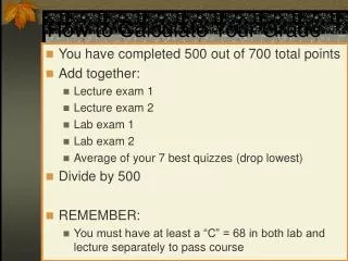
How to Calculate Your Grade • You have completed 500 out of 700 total points • Add together: • Lecture exam 1 • Lecture exam 2 • Lab exam 1 • Lab exam 2 • Average of your 7 best quizzes (drop lowest) • Divide by 500 • REMEMBER: • You must have at least a “C” = 68 in both lab and lecture separately to pass course
How to Calculate the Number of points you need to pass • You need a total of 476 out of 700 points to get a C • Take the total number of points you just calculated (the sum of 4 exams and quiz average) and subtract it from 476 • The number you have is the total number of points you need • If you divide that number by 2, you will see the approximate grade you’ll need on lab exam 3 and lecture exam 3. • REMEMBER: the rules from previous page apply • Have to have at least C in lecture and lab separately
Organs of the Abdomen Systems: Urinary and Digestive
Urinary System • Kidneys • Purify blood • Ureters • Drain urine from kidney to bladder • Urinary Bladder • Store urine • Urethra • Drain urine from bladder to outside body pg 5
Kidneys: major excretory organs • Remove toxins, metabolic waste, excess H2O, ions • Urea, uric acid, creatinin • Regulates volume + makeup of blood • Maintains balance between • Salts and water • Acids and bases
Kidneys: Gross Anatomy • Located superior lumbar region • Posterior abdominal wall (T12-L3) • Retroperitoneal • Hilus • Adrenal Gland: superomedial to kidney • Renal Artery + Vein • Innervation: branches of renal plexus pg 648
Kidneys: Gross Anatomy • Renal Capsule • Layer of tough CT • Maintains shape • Prevents spread of infection • Adipose Capsule • External to renal cap • Perirenal fat • Surrounded by fascia • Keeps in place, cushions • Pararenal Fat • External to adipose cap • Keeps in place, cushions pg 649
Kidney: Internal Anatomy • Cortex • Superficial • Light, granular • Part of functional unit • Medulla • Deep layer • Darker • Pyramid-cone shape • Contain collecting tubule collect urine Pg 650
Kidney: Internal Anatomy • Medullary Pyramid • Base: against cortex • Apex: inward • Papilla = tip • Drips urine into minor calyx • Minor Calyx (calices) • Cup-shaped divisions of major calices • Surround papilla of pyramid • Major Calyx (calices) • Larger cup-shaped branches of renal pelvis • Renal Pelvis • Flat expansion of ureter • Collects urine Pg 650
Kidney: Microscopic Anatomy • Functional Unit • Uriniferous Tubule • Nephron • Collecting tubule • Waste is filtered out • Waste products formed • Located in lobes of kidneys pg 652
Ureters • Slender tubes transport urine • Run from kidneys to bladder • Retroperitoneal • Continuation of renal pelvis • Enters bladder at oblique angle to prevent backflow • Increased pressure in bladder closes distal end of ureter pg 648
Ureters: 3 Layers • External: Adventitia • CT • Middle: Muscularis • Smooth Muscle • Inner Longitudinal • Outer Circular • External longitudinal (on distal third) • Peristalsis • Inner: Mucosa • Transitional epithelium
Bladder • Muscular sac store and expel urine • Location • On pelvic floor • Posterior • Pubic symphysis • Anterior • Males = rectum • Females = vagina, uterus • Collapses + Expands • Full into abdominal cav • Emptystays in pelvic cav • Supplied by branches of internal iliac arteries + veins • Innervated = branches of hypogastric plexus pg 648
Bladder: Internal Anatomy • 3 Layers • Mucosa = transitional epithelium & lamina propria • Detrusor Muscle: smooth muscle • Inner/Outer longitudinal, Middle circular • Fibrous Adventitia = CT • Parietal peritoneum on superior surface instead trigone pg 662
Urethra • Drains urine from bladder to outside • Female = short tube • Males = long tube • Prostatic, Membranous, Spongy (penile) portions • Also carries semen • Internal Urethral Sphincter • Between bladder + urethra • Thickening of detrusor (smooth muscle) • External Urethral Sphincter • Within urogenital diaphragm • Skeletal muscle = voluntary control urination • External Urethral Orifice • Males = end of penile urethra • Females = anterior to vaginal opening, posterior to clitoris
Urethra: Female vs. Male pg 662
Micturition = Urination • Emptying bladder • Stretch receptors in bladder respond when bladder full • Parasympathetic signals detrusor muscle to contract and internal urinary sphincter to open (also inhibits sympathetic pathways that would prevent urination) • Other brain receptors can inhibit urination by relaxing detrusor, and keep external urinary sphincter closed • Voluntary contraction of abdominal wall muscles increases abdominal pressure • Voluntary relaxation of external urethral sphincter See pg 663
Digestion System • Alimentary Canal • Mouth • Pharynx • Esophagus • Stomach • Small Intestine • Large Intestine • Accessory Organs • Teeth, Tongue • Salivary Glands • Gallbladder • Liver • Pancreas pg 5
Food Processing Activities • Ingestion: taking food into mouth • Propulsion: food moves through gut • Swallowing + Peristalsis • Mechanical Digestion: breakdown of food • Chewing, Churning, Segmentation • Chemical Digestion: chemical breakdown • Enzymes • Absorption: Digestive end products into blood • Defecation: Removal of waste products
Alimentary Canal Wall • Internal = Mucosa + Submucosa • Epithelium • Lamina propria: • contains MALT: mucosa-associated lymphoid tissue • Muscularis mucosae • Submucosa = CT w/elastic fibers, nerves, vessels • Middle = Muscularis Externa • Inner circular layer • Outer longitudinal layer • Creates sphincters • Outer = Serosa or Adventitia
Innervation of Alimentary Canal • 2 Plexuses: Myenteric & Submucosal • Parasympathetic, Sympathetic, Visceral Sensory fibers • Enteric Nervous System • 100 million neurons in walls of alimentary canal = internal system • Within above plexuses • Independent reflex arcs • Controls glandular secretion, peristalsis, segmentation • Autonomic Nervous System speeds up or slows activity controlled by enteric system
Stomach • “J” shape • Cardiac Region • Junction esophagus • Cardiac sphincter (Gastroesophageal) • Fundus (“dome”) • Under diaphragm • Body • Large, middle part • Pylorus • Distal portion • Pyloric sphincter • Greater Curvature • Lesser Curvature Pg 624
Internal Anatomy of Stomach • Mucosa • Rugae: mucosal folds allow expansion • Many intrinsic glands • Goblet cells • Gastric glands • Typical Submucosa • Muscularis externa • Oblique layer • Circular layer • Pyloric sphincter • Longitudinal layer • Serosa pg 624
Function of Stomach • Temporary storage of chyme • Breakdown begins • Churn, segmentation • Pepsin proteins • Absorption • H2O, electrolytes • Alcohol, other drugs • Stays about 4 hours • Hold from1.5-4 liters
Small Intestine: Parts + Functions • Parts • Duodenum = proximal (5%) • Jejunum = middle (~40%) • Ileum = distal (~55%) • Majority of enzymatic digestion • Bile: emulsifier (gallbladder, liver) • Enzymes (pancreas) • Almost all nutrient absorption • Segmentation • Moves chyme around to increase contact with intestine walls • Food takes about 3-6 hours to move through • 2.7- 6 meters
Small Intestine: Internal Features • Intestinal flora: produce vitamin K • Simple columnar epithelium w/many modifications for absorption • Lymph tissue in submucosa • Muscularis externa has 2 layers • Some parasympathetic innervation from vagus • Arterial supply: • Superior mesenteric • Rt (cranial) pancreaticoduodenal
Small Intestine:Modifications of epithelium for absorption • Length • Increase surface area • Plicae circularis • Transverse ridges of mucosa • Increase surface area • Slow movement of chyme • Villi • Move chyme, increase contact • Contain lacteals: remove fat • Microvilli: • Increase surface area • Modifications decrease distally pg 629
Small Intestine • Duodenum: • short, straight • Mostly retroperitoneal • Jejunum & Ileum: • highly coiled • Fewer modifications • Hang by mesentery in peritoneal cavity • Mesentery Arcades • Arteries + veins • Nerves • Store fat Pg 614
Large Intestine • Cecum • Vermiform appendix • Colon • Ascending • Transverse • Descending • Sigmoid • Rectum • Anal Canal pg 631
Large Intestine • Functions: • Absorb water and electrolytes • Form, store and expel feces from body • Internal Features: • Intestinal flora • No intestinal villi or modifications for absorption • Many goblet cells • Simple columnar epithelium except lower half of anal canal • Significant Lymph tissue in mucosa & submucosa • Muscularis mucosae has 2 layers • Some parasympathetic innervation from vagus
Colon: External Features • Taeniae coli • 3 longitudinal strips • thickening of longitudinal muscle • maintain muscle tone • create haustra • Haustra • saclike divisions • Epiploic Appendages • fat-filled pouches • significance unknown pg 631
Cecum + Vermiform Appendix • Cecum • sac-like, blind pouch • Ileocecal valve • raised edges of mucosa • prevents feces going back into ileum • Vermiform Appendix • same layers • blind tube opens into cecum • masses of lymph tissue pg 631
Colon • Ascending colon • Right side • Hepatic flexure (= right colic flexure) • Transverse colon • Across cavity • Descending colon • Left side • Splenic flexure (= left colic flexure ) • Sigmoid colon • Enters pelvis • “S” shape pg 631
Colon: Function • Absorb H2O and electrolytes • Some digestion by bacteria • Mass Peristaltic Movements (2-3x day) • Moves through in 12-24 hours • 1.5 meters
Rectum + Anal Canal • Rectum • descends into pelvis • no teniae coli • longitudinal muscle layer complete • rectal valves • Anal Canal • passes through levator ani muscle • releases mucus to lubricate feces • Internal anal sphincter • involuntary, smooth m. • External anal sphincter • voluntary, skeletal m. • Stratified squamosal epithelium at lower half pg 632
Defecation Reflex • Stretching of rectum wall initiates reflex • Spinal cord - parasympathetic signals sigmoid colon + rectum to contract + anal sphincter to relax (involuntary) • If not ready-reflex ends- rectum relaxes • Reflex initiated again until you go! • Contraction of abdominal muscles, levator ani + diaphragm assists defecation (voluntary)
pg 610 Liver • Largest gland (3 lbs) • Location • Upper Right Quadrant • Mostly under ribcage • Highly vascular • Some functions • produce bile • pick up glucose • detoxify poison, drugs • make blood proteins • many others pg 635
Liver: External Features • Diaphragmatic surface • Right lobe (larger) • Left lobe • Falciform ligament • Fissure between • Visceral surface • Quadrate lobe • Caudate lobe • Both part of left lobe pg 635
Liver: Visceral Surface • Hepatic Vein (into inferior vena cava) • Porta Hepatis • Hepatic Artery (from abdominal aorta ) • Hepatic Portal Vein • Carries nutrient-rich blood from stomach + intestines to liver • Portal system = 2 capillary beds! • Hepatic Ducts (carry bile) pg 636
Gallbladder • Muscular sac • Between right + quadrate liver lobes • Bile is stored + concentrated • Bile: breaks down fats = emulsification • Bile • Produced by liver • Stored in gallbladder pg 610
Gallbladder continued • Mucosa & lamina propria • Simple columnar epithelium • Expandable mucosal folds • Smooth muscle layer • Thick connective tissue • Covered by serosa in places
Bile Ducts • Cystic duct • carries bile from gallbladder • Hepatic duct • carries bile from liver • Common Bile duct • joins cystic and hepatic • carries bile into duodenum pg 628
Movement of Bile • Bile secreted by liver continuously • Hepatopancreatic (Vater) ampulla • common bile + main pancreatic duct meet and enter duodenum • Sphincter of Oddi around it • closed when bile not needed for digestion • Bile then backs up into gallbladder via cystic duct • When needed gallbladder contracts, sphincters open pg 628
Pancreas • Retroperitoneal • Gland • Exocrine • digestive enzymes • Endocrine • hormone insulin • hormone glucagon • Location • curve of duodenum • extends to spleen pg 639
Ducts of Pancreas • Main Pancreatic duct • joins common bile duct • enters duodenum • Hepatopancreatic (Vater) ampulla • Accessory Pancreatic duct • enters duodenum in other location pg 628
Spleen • Largest lymph organ • Highly vascular • Function • remove blood-borne antigens (immune) • remove and destroy old/damaged blood cells • stores blood platelets • In fetus: site of hematopoiesis pg 639
Arterial Blood Supply to Abdominal Viscera • All branches of Abdominal Aorta • Anastomoses • Left + Middle colic • Left + Right gastric • Left + Right gastroepiploic • Cranial + Caudal pancreaticoduodenal • Deep Iliac Circumflex + Adrenolumbar • STUDY HAND OUT! MUST KNOW WHAT SUPPLIES WHAT!!
Hepato = liver Pancreatico = pancreas Cystic = gallbladder Gastro = stomach Splenic = spleen Adreno = adrenal gl Lumbar = lumbar region Epiploic = membrane-covered Mesenteric = mesentery Duodenal = duodenum Ileo = ileum Colic = colon Rectal = rectum Names give hints!