Download
1 / 11
110 likes | 269 Vues
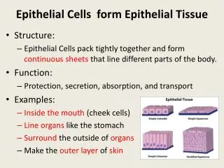
Effects of smoking in human lens epithelial cells. Jae-Woong Koh (MD/PhD) 1 , Nang-Hee Song(MD) 1 , Gil-Joong Yoon (MD/PhD) 2 Department of Ophthalmology, Chosun University College of Medicine, Gwangju, Republic of Korea 1 Happy Eye Clinic, Gwangju, Republic of Korea 2

E N D
Effects of smoking in human lens epithelial cells Jae-Woong Koh (MD/PhD)1, Nang-Hee Song(MD)1, Gil-Joong Yoon (MD/PhD)2 Department of Ophthalmology, Chosun University College of Medicine, Gwangju, Republic of Korea1 Happy Eye Clinic, Gwangju, Republic of Korea2 Authors have no financial interest.
Dr. Koh Dr. Song Dr. Yoon
Introduction Cadmium • A highly toxic chemical material • Group I carcinogen in humans by the international agency for Research on Cancer (IARC, 1993) • Two major source (Cigarette smoking, Food because of its high rate of soil to plant transfer) • Accumulated in human bodies because of the Long biological half–life(10-30yrs) • Accumulates in various ocular tissues such as lens, retina, ciliary body and vitreous. Cadmium & Cataract • Elevated cadmium has been reported in cataractous lenses compared to clear human Lenses (Ramakrishnan et al., 1995; cekic, 1998) • A significant amount of cadmium in the lenses of smokers of chronic smokers (Mosad et al., 2010:Ramakrishnan et al. , 2010) • The smokers exhibit early cataract formation (Claytin et al., 1984)
Introduction P53 • The tumor suppressor gene is activated upon DNA damage and it is accumulated in the nucleus, where it then functions as transcription regulator of for its downstream target gene. Caspases • A family of cysteine–dependent aspartate directed proteases, play critical role in the initiation and execution of apoptosis. • Two major pathway for caspases activation (extrinsic and intrinsic path ways) (1) The extrinsic pathway involves the ligation of death receptors resulting in caspase-8 activation. This initiator caspases activates other caspases (Caspase 3, 7) referred to as effector caspases. (2) The intrinsic pathway, In the cytosol, cytochrome c binds to and activates Apaf-1 which itself activates pro caspase-9. Activated caspase- 9 has been shown to directly cleave and activated the effector protease, caspase-3.
Purpose/Method Purpose • To investigate the cellular change and cell death mechanism in cultured human lens epithelial cells induced by smoking. Method • Cultured lens epithelial cells were challenged with 200, 400, 600, 800, 1000 uM of cadmium chloride (CdCl2, Catalog No. 202908, Sigma-Aldrich Chemical Co, USA) for 2 hours, and then were exposed to UV (280-320nm/ 1.2mv/cm2) • The cell viability was evaluated using the microscope and 3-(4,5-dimethylthiazol -2-yl)- 2,5-dipheny tetrazolium bromice (MTT) assay. • The gene and protein level of caspase-8 and P53 were measured after exposed to CdCl2(600uM) for 2 hours by using RT PCR and western blot.
Result • Compared to untreated cell, the cell death increased after Cadmium chloride exposure in microscopic finding. And MTT assay demonstrated that the cell death was increased in proportional to the increased Cadmium chlorideconcentration. • The expression of caspase-8 and P53 level all increased after exposure to cadmium (600 uM ) in RT PCR and western blot.
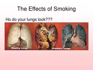
Result Photograph of control cells and primary bovine lens epithelial cells exposed to 800 uM cadmium chloride for 4 hours. Apoptotic features such as shrinkage of cytoplasm and nuclear fragmentation were better observed in the cadmium treated group than control group. Photographed using phase contrast microscopy. Magnification x 100 Control (100X) 4 hr after CdCl2(800uM) treatment (100X)
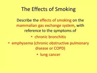
Result Effect of cadmium on the viability of HLCEs Dose dependent declined in HLECs viability by cadmium. Serum starved HLECs were incubated with cadmium (0-1000uM) for 24 hr followed by determination of cell viability by MTT assay (Mean ± SD of 3 experiments)
Result Western blot and Quantitative analysis shows that production of caspase 8 protein in those cell in Increased after Exposure to 600 uM cadmium. Western blot and Quantitative analysis shows that production of P53 protein in those cell in increased after Exposure to 600 uM cadmium. Caspase 8 P53 Control P53 Control caspase 8
Conclusion Our study has successfully shown • Cadmium causes a significant decline in the viability of HLECs in a dose dependent manner. • Cadmium-induced decrease in cell viability is due to apoptosisin HLECs. • Cadmium-induced apoptosis occurred via the activation of caspases.
Reference 1) Ramkrishnan. S. Sulochana, K.N. Selvaraj. T et al, smolking of beedied and ataract : camium and vitamin C in the lens and blood. Br J ophthalmol 79;202-206 2) Mosad. S.M Chanem A.A El-Fallai et all, Lens cadmium lead and serum vitamin C, E and beta carotene in cataractous smoking patient. Curr eye res 201035:23-30 3) Clayton R.M, Cuthbert J et all Epidemiological and other studies in the assessment of factors cotributing to cataractogenesis. Ciba Found symp 1984;106;25-47 4) Johar, S.R., Rawal, U.M., Jain, N.K., Vasavada, A.R., 2003. Sequential effects of ultraviolet radiation on the histomorphology, cell density and antioxidative status of the lens epithelium: an in vivo study. Photochem. Photobiol. 78, 306–311. 5) Li, W.C., Kuszak, J.R., Dunn, K., Wang, R.R., et al., 1995. Lens epithelial cell apoptosis appears to be a common cellular basis for non-congenital cataract development in humans and animals. J. Cell Biol. 130, 169–181. 6) Spector, A., 1995. Oxidative stress induced cataract: mechanism of action. FASEB J. 9, 1173–82 7) Takamura, Y., Kubo, E., Tsuzuki, S., Akagi, Y., 2003. Apoptotic cell death in the lens epithelium of rat sugar cataract. Exp. Eye Res. 77, 51–57 8) Kim, S., Moon, C., Eun, S., Ryu, P., Jo, S., 2005. Identification of ASK1, MKK4, JNK, c-Jun, and caspase-3 as a signaling cascade involved in cadmium-induced neuronal cell apoptosis. Biochem. Biophys. Res. Commun. 328, 326–334. 9) Chen, L., Liu, L., Huang, S., 2008b. Cadmium activates the mitogen-activated protein kinase (MAPK) pathway via induction of reactive oxygen species and inhibition of protein phosphatases 2A and 5. Free Radic. Biol. Med. 45, 1035– 44 10) Nelesh M. Kalariya, Bindu Nair, Denish K. Kalariya, Nancy K willis et al, 2010, Cadmium- induction of cell death in human lens epithelail cells : Implication to smoking associated cataractogenesis. Toxicoloy letters198;56-62