Download
1 / 41
1.16k likes | 4.74k Vues
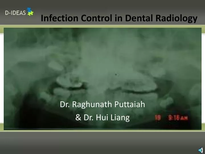
Infection Control in Dental Radiology. Dr. Raghunath Puttaiah & Dr. Hui Liang. Introduction & Rationale. Most oral and maxillofacial radiology normally consists of non-invasive procedures Although exposure to blood is not common, contact with saliva does occur. What are the risks??.

E N D
Infection Control in Dental Radiology Dr. Raghunath Puttaiah & Dr. Hui Liang
Introduction & Rationale • Most oral and maxillofacial radiology normally consists of non-invasive procedures • Although exposure to blood is not common, contact with saliva does occur
What are the risks?? • Common viruses seen in the oral cavity include— • cytomegalovirus, herpes simplex viruses-1 & 2, hepatitis-B and D viruses, hepatitis-C virus, influenza viruses, Epstein-Barr virus (infectious mononucleosis), Rhinoviruses (common cold), and HIV • Common bacterial pathogens seen are— • Pseudomonas, Flavobacterium, Staphylococci, Streptococci, Diplococci, Pneumococci, Mycobacterium, Chlamydia, and Spirochetes from human or inanimate sources • Candidiasis is also very common among dental patients
Rationale • Most of Oral and Maxillofacial radiology (OMR) procedures fall mainly in the semicritical and non-critical categories of Spaulding's Classification of inanimate surfaces BUT • Many contagious diseases such as— • Infectious mononucleosis and hepatitis-B can possibly be spread by simple contact with saliva • Therefore, it is necessary for aseptic techniques to reduce ambiguity within protocols • The purpose of this module is to present the most recent information regarding aseptic techniques in OMR.
What PPE is needed • It is not usually necessary to wear PPE such as impervious gowns, long sleeves, masks and protective eyewear during routine OMR procedures when no aerosols, droplets or spatter are generated • One may use gloves, gowns, masks and protective eyewear while treating patients with gagging problems • While handling the processor and the chemicals, use full PPE • Stay out of the Radiation Hazard area (behind a shield) • Patients must be protected with a lead apron
Disinfectants and Barriers • Barriered films should be used when available • Between patients, frequently touched areas must be— • Barriered with plastic barriers OR -Disinfected with an Intermediate level, Hospital Disinfectant using a Spray-Wipe-Spray technique • If barriers are used between patients, disinfection is only needed at the beginning or end of the day
Steps in Infection Control Unit Dose— 1. Pre-procedural mouthrinse for patients Make them rinse for about 30 seconds to reduce the microbes in the mouth 2. PPE- Gloves Masks Eyewear Gown
Steps in Infection Control Prepackaged films with plastic barriers
Steps in Infection Control • Unit dose supplies and sterile equipment on to a clean bib/work • surface • 2. Set up the films on to the positioning devices • 3. Label cups as Exposed and Unexposed
Use of Disinfectants and Barriers • Disinfect surfaces at the beginning and end of each day, and between patients only if contaminated • Avoid spraying electrical switches, wipe with disinfectant moistened paper towel • Apply surface covers to the yoke, tube head, cone, control unit, head rest, arm rest, and any hand held switches
Aseptic Procedures • Place the sanitized lead apron and collar on the patient after seating the patient • Set the required mA, kVp and exposure time on the control unit and reset as required • Use Foot Controlled Trigger Switch if possible
Now Please expose the Radiograph The whole purpose in dental radiology is to Get the radiograph
Inside a daylight loader • Place the cup with exposed films on one side and the • Empty one on the other • Place a clean bib or napkin as a barrier • Dispense a pair of gloves
Daylight Loader with Films Inside • Be sure to close the lid before exposing the films
Various layers inside the polysoft film cover Polysoft cover Radio-opaque metal foil on the rear of the film Film Paper folder that secures the film
Processing Barriered Films Exposed film • Wear gloves • Peel the outer cover and drop • the film into the “Exposed” cup • This should be done chairside
Step 1 Step 2 Step 4 Step 3
Unbarriered Films Step 2 Step 1 Step 3 Step 4
Bare hands to be used tohandle exposed and peeled film Remove your gloves and then handle the film that has been taken out of the Polysoft cover
Clean up after loading films • Carefully hold the edge of the napkin or bib and fold it over the waste • You need not wear gloves if you hold the non-contaminated corners
Disinfect the Daylight Loaderonce or twice daily if barriers are used Barriers limit the use of disinfectants
Panoramic Radiography • In panoramic radiography, infection control procedures are very simple • Patient needs to have a lead apron • No contact of film with saliva • Only one barrier is needed
Panoramic Radiography Barriered positioning device Bite block Chin rest
Panoramic Radiography • Infection Control in Panoramic • radiography is very simple • All one needs is a barrier for • the bite block • Make sure the lead apron is on • Film loaded on to the cassette • Stay outside of the active • radiation area
Panoramic Radiography This film does not come in contact with saliva
Panoramic Radiography Position the patient correctly
Panoramic Radiography Make the patient remove the barrier on the bite block
Digital Radiography Sensor being inserted into barrier Sensor Sensor Barriered Barrier or sleeve
Digital Radiography • Insert the barriered sensor in the film holder on the Rinn holder
Digital Radiography Slide this door open
Digital Radiography Peel open the barrier without touching the sensor
Digital Radiography Slide the sensor into the chamber for the computer to read the image
Digital Radiography Image appears on the screen within seconds