Download

1 / 21
210 likes | 331 Vues
XIX Symposium Neuroradiologicum. Perifocal MR Perfusion and Diffusion Values in Gliomas. Z. Rumboldt, M.V. Spampinato, P. Morgan, C. Schiarelli, C. Rorden, J. Fridriksson . Medical University of South Carolina Charleston, SC, USA. Background.

E N D
XIX Symposium Neuroradiologicum Perifocal MR Perfusion and Diffusion Values in Gliomas Z. Rumboldt, M.V. Spampinato, P. Morgan, C. Schiarelli, C. Rorden, J. Fridriksson Medical University of South Carolina Charleston, SC, USA
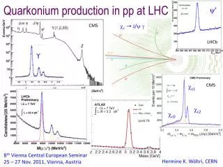
Background New therapies for gliomas highlight thelimits of conventional imaging as a tool to assess tumor extension and progression Growing interest in advanced MR techniques such as perfusion and diffusion imaging to develop better ways to assess treatment response and prognosis Henson et al. AJNR Am J Neuroradiol. 2008 Mar; 29(3):419-24. Dhermain FG et. Lancet Neurol. 2010 Sep;9(9):906-20
MR Perfusion High grade gliomas characterized by increased neovascularization rCBV is a parameter correlating with the amount of capillaries within the tumor Aronen, Neuroimaging Clin N Am. 2002 Nov;12(4):501-23
ADC Role of diffusion-weighted MRI in grading gliomas Pre- and post-treatment DWI data could assist in predicting response to anti-VEFG Arvinda HR et Al. J Neurooncol 2009; 94: 87–96 Pope WB et Al. Radiology 2009; 252: 182–89 Wu J et Al. J Clin Oncol 2010; 28 (suppl)
Purpose Validation Study Hypothesis: Measurements of rCBV and ADC within the tissue beyond the abnormality seen on conventional MR imaging may provide relevant information regarding the biological behavior of cerebral gliomas These data could help predicting the aggressiveness of a neoplasm, determining treatment response, and establishing prognosis
Materials and Methods • 11 patients with cerebral glioma • (5 WHO grade IV, 1 grade III, 5 grade II) • – 6 progressed in 6 mos • - 5 stable for 2 years • - 4 oligodendroglial (3 low, 1 high grade) • - 1.5T MR scanners (pre or post treatment) • - ADC • - rCBV
Materials and Methods • rCBV analyzed off-line using the Java • Image software package (www.xinapse.com) • - Deconvolution of the arterial input function from the tissue response function according to the method described by Ostergaard et al. Ostergaard et Al. Magn. Reson. Med. 36:715-728 (1996).
Materials and Methods • JIM software rCBV maps and rCBV maps processed usingdifferent vendors’ software were compared • Image quality was rated in consensus by two neuroradiologists (random order, using a 3-point scale)
Materials and Methods • -Tumor delineation: VOIs were drawn on all relevant FLAIR images using MRIcron (http://www.cabiatl.com/mricro/mricron) • VOI overlaid on the ADC and rCBV maps
Materials and Methods • In addition to the lesion VOI, mean values • also obtained from incrementally dilated regions • automatically defined using MRIcron software • Dilationbands in 3D ranges away from • the lesion at predefined distances: • 0-4mm, 5-9mm, 10-14mm, 15-19mm • Dilation constrained to ipsilateral hemisphere • Ventricles excluded
Materials and Methods • Statistical Analysis • Kruskal Wallis • Grouping variable: progression • rCBV • ADC absolute values
Results rCBV images processed by Java Image software package: - superior in 6 cases - equal in 5 cases
Results - rCBV Progression Stable
Results - ADC Progression Stable Perifocal ADC values Dropping
Conclusion Java Image (JIM) software package can be used for brain MR perfusion analysis MRIcron may be a powerful tool for evaluation of perifocal tissue in brain gliomas ADC is routinely obtained, no postprocessing needed Perifocal absolute ADC values appear to discriminate as well as rCBV
Conclusion rCBV and ADC values in perifocal dilated regions may provide additional information that can assist in tissue characterization Further prospective investigations are necessary May make substantial contributions to treatment response and prognosis prediction