Download
1 / 42
420 likes | 437 Vues
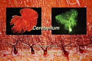
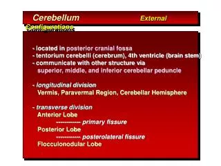
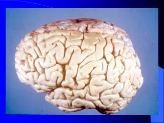
Cerebellum: Anterior Aspect. Lateral hemisphere. Vermis. Posterior Lobe. Primary Fissure. Anterior Lobe. The cerebellum has three lobes: anterior , posterior , and flocculonodular . Each lobe has a vermal portion and a hemispheric portion. Midbrain. Cerebellum, Posterior View.

E N D
Cerebellum: Anterior Aspect Lateral hemisphere Vermis Posterior Lobe Primary Fissure Anterior Lobe The cerebellum has three lobes: anterior, posterior, and flocculonodular. Each lobe has a vermal portion and a hemispheric portion. Midbrain
Cerebellum, Posterior View Vermis Pyramis Posterior LobeHemisphere Uvula Tonsils of cerebellum Medulla Foramen of Magendie (opening in post. medullary velum from 4th ventricle)
The flocculus is the hemispheric part of the flocculonodular lobe of the cerebellum. The nodule is the vermal part of the flocculonodular lobe. Primary fissure Ant Post Tonsil Prenodular fissure Posterolateral fissure
Cerebellar Vermis The cerebellar vermis has ten sublobules. Primary fissure Midbrain Anterior lobe vernis Fourth ventricle Pons Posterior lobe vermis Nodule Prenodular fissure Medulla Tonsil
Cerebellar Peduncles: connect the cerebellum to the brainstem; each pair of peduncles connects the cerebellum to a separate division of the brainstem: SCP to midbrain, MCP to pons, and ICP to medulla. Inferior cerebellar peduncle- afferents (input) spinocerebellars, cuneocerebellars, vestibulocerebellars, and olivocerebellars Middle cerebellar peduncle- afferent, pontocerebellars Superior cerebellar peduncle- primarily efferent (output) cerebellorubrals (to red nucleus) and cerebellothalamics (to VL nucleus of thalamus)
The cerebellar cortex contains three layers: molecular, Purkinje cell, and granular layers Molecular layer Cerebellum Purkinje cell layer Cell body of Purkinje cell Granular layer Primary dendrite Vermis Abundant dendritic spines on dendrites Dendrites Cell body of Purkinje neuron H & E Cerebellar Cortex Axon
Intrinsic Cerebellar Cortex Connections Granule cells give rise to parallel fibers Purkinje cells Mossy fibers terminate on granule cells Purkinje cells project to deep cerebellar nuclei Climbing fibers terminate on Purkinje cells
Vermis Paravermal cortex Purkinje cells of the cerebellar cortex project in topographic order to deep cerebellar nuclei. Cerebellar efferents from the dentate, globose, and emboliform nuclei join the SCP to project to the contralateral (CL) red nucleus and VL nucleus of the thalamus. The fastigial nucleus has reciprocal connections with the vestibular complex thru the juxtarestiform body. Lateral Hemisphere E G F D SCP JRB CL Red nucleus VL Nucleus Vestibular complex Inferior olivary nucleus Spinal cord Motor cortex
Cerebellar Afferents: Spinocerebellars Cuneocerebellars Vestibulocerebellars Pontocerebellars Olivocerebellars All of these inputs enter the cerebellum thru the inferior & middle cerebellar peduncles. All (except olivocerebellars) terminate as “mossy fibers” on granule cells. Olivocerebellars terminate as “climbing fibers” directly on Purkinje cells.
Cerebellar Afferents Spinocerebellar and cuneocerebellar tracts primarily terminate in the anterior lobe Pontocerebellars terminate in the posterior lobe Olivocerebellars go to all lobes Vestibulocerebellars terminate in the flocculonodular lobe
Pontocerebellar Connections relay information from motor cortex about ongoing movements Corticopontine tract Posterior lobe (neocerebellum) Basilar pontine nuclei Pontocerebellar fibers terminate primarily in the posterior lobe. Olivocerebellar fibers terminate in all lobes
Vermis Paravermal cortex Purkinje cells of the cerebellar cortex project in topographic order to deep cerebellar nuclei. Cerebellar efferents from the dentate, globose, and emboliform nuclei join the SCP to project to the contralateral (CL) red nucleus and VL nucleus of the thalamus. The fastigial nucleus has reciprocal connections with the vestibular complex thru the juxtarestiform body. Lateral Hemisphere E G F D SCP JRB CL Red nucleus VL Nucleus Vestibular complex Inferior olivary nucleus Spinal cord Motor cortex
Cerebellar Connections: Principle of Lateralization Spinocerebellar and Vestibulocerebellar Inputs are primarily ipsilateral (conveying information about the same side of the body) Cerebellar efferents project to the contralateral VL nucleus of the thalamus and motor cortex. Corticopontines project to basilar pons, and pontocerebellar afferents cross back to the original side. Hence, clinical deficits resulting from cerebellar lesions are expressed ipsilaterally.
Cerebellar Lesions: deficits expressed ipsilaterally Ataxia- tendency to fall toward side of lesion IntentionTremor (Action Tremor) Dysdiadokinesia- inability to produce alternating antagonistic actions Past-pointing Nystagmus- flocculonodular lobe lesion