Download
1 / 76
760 likes | 896 Vues
Bio 211- Anatomy and Physiology I . Today’s topics Tissues. Structural hierarchy in human biology. Biggest, most complex. Human Body Organ system (cardiovascular system) Organ (Heart) Tissue (cardiac muscle) Cells ( cardiomyocytes ) Organelles (nucleus, ribosome)

E N D
Bio 211- Anatomy and Physiology I • Today’s topics • Tissues
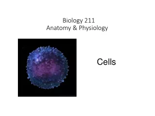
Structural hierarchy in human biology Biggest, most complex Human Body Organ system (cardiovascular system) Organ (Heart) Tissue (cardiac muscle) Cells (cardiomyocytes) Organelles (nucleus, ribosome) Macromolecule (Protein, DNA) Molecule (amino acid) Atom (nitrogen, carbon, oxygen) Smallest DONE!!
Histology • The study of cells and tissues and how they are arranged into organs • Allows us to see how cells are arranged in a particular tissue type and how tissues help form an organ • 200+ types of cells in our bodies are broken down into 4 primary tissue classes or categories: • Epithelial • Connective • Nervous • Muscular
Tissues • A group of similar cells derived from a common embryonic origin that are arranged in a way that allows them to carry out a particular STRUCTURAL or PHYSIOLOGICAL function • STRUCTURAL • Bone (type of connective tissue) supports and protects the body and allows muscles to produce movement • PHYSIOLOGICAL • Epithelial tissues lining the digestive system allows the body to absorb nutrients from the foods we eat • Many tissues have both a structural and physiological function (bone) Tissue = cells + extracellular matrix (or extracellular material) • Tissues differ from one another in the types of cells which make up the tissue, as well as by the type and amount of EXTRACELLULAR MATRIX that surrounds the cells • Matrix contains protein fibers, water, minerals, nutrients, waste products
Studying Histology Biologists use microscopes to examine tissue sections (very thin slices) colored with various stains or antibodies (used to visualize proteins) to determine the location and arrangement of cells within a tissue Depending on the stain used, you can visualize cytoplasm, nuclei, even specific proteins! Purple nuclei Brown stain shows location of a specific lung protein Pink cytoplasm
Epithelial Tissues • EPITHELIAL TISSUES are characterized by a layer of tightly bound cells that are one or more layers thick (1-20), bound to a BASEMENT MEMBRANE and are usually exposed to the outside environment or to the inside of the body • Examples : skin, lining of digestive system, lining of lungs, lining of blood vessels, etc… Epithelial cells Basement membrane Connective tissue • Basement membrane acts like a biological glue to hold cells to connective tissue underneath • composed of collagen, and a variety of glycoproteins and proteoglycans
Classification of epithelial tissues • Cell Shapes • Squamous – thin, flat • Good for exchange of gases, nutrients, waste products (lung and blood vessels) • Cuboidal – typically square or round • Found lining the ducts of many glands • Columnar – tall and narrow • Found lining the intestines • Often have small projections on exposed surface (MICROVILLI) that increase surface area for absorption
Classification of epithelial tissues • Simple – have a single layer of cells • Stratified – Tissue contains a few to many layers of cells on top of each other • Only bottom layer are bound to basement membrane • Upper layers have cells bound to other cells • Transitional – Specialized type of epithelia found in organs that stretch • Pseudostratifiedcolumnar – all cells are bound to the basement membrane but not all cells reach the exposed surface.
Epithelial tissue histology Simple cuboidal: Single layer of cube or round cells Simple Squamous: Single, layer of thin cells
Pseudostratified columnar: Single layer of columnar cells, nuclei are scattered, not all cells reach exposed surface Simple columnar: Single layer of tall cells, all cells reach the exposed surface, microvilli present Microvilli
Keratinized, stratified squamous epithelia: Many layers of cells, lower cells are alive and dividing, upper cells are dead and filled with keratin, CANNOT SEE NUCLEI Non-keratinized stratified squamous epithelia: Many layers of cells, all alive, CAN SEE NUCLEI
Transitional epithelium: • Cells are alive and overlapping, may appear single layer when tissue is stretched • Found in tissues that need to stretch on a regular basis • Have a cuboidal appearance in a relaxed tissue but “TRANSITION” to a squamous appearance when stretched
Tips to identifying and classifying epithelial tissues • Try to identify how many layers of cells are between the basement membrane (bottom) and the exposed surface. Living cells will have visible nuclei, but dead keratinized cells will not. • Try to identify the shape – thin, flat (squamous), cube or round (cuboidal), tall and thin (columnar) • Number of layers and shape of cell tells you the name of the tissue • Stratified sqaumous (SEVERAL LAYERS of THIN, FLAT cells) • Simple cuboidal(ONE LAYER of CUBE-SHAPED cells) • Etc…. Since epithelial tissues are often found lining something you will typically see epithelial tissues next to open spaces (white space on a microscope slide)
Connective Tissue Tissue derived from the mesoderm that provides protection and support for the body and its organs, often composed of dense fibers, large amount of extracellular material with relatively few cells • Major Functions: • Connection – connective tissue binds bones to muscle (tendons), bones to other bones (ligaments) and keeps organs in place • Support – bones help support the weight of the body, cartilage supports structures like the nose and ear • Protection – • Physical- delicate organs like the brain, kidneys, eyes, etc… are often surrounded by bone and adipose tissue for protection • Immune – LEUKOCYTES (white blood cells) and LYMPHATIC organs are connective tissues that protects the body from invasion by bacteria and viruses • Movement – Bones help move body with help of muscle, muscles attached to bones compress some body cavities • Storage and heat production – Adipose tissue (Fat) stores energy for later use and preserves/generates heat • Transport – Blood transports gases, nutrients, waste, hormones to distant parts of the body
Types of connective tissue • Fibrous connective tissue – • What most people think of when talking about connective tissue • Ligamentsand tendons contain fibrous C.T. • Cartilage – • Rubbery C.T. that supports nose and ears, found between bones • Bone – • Rigid C.T. that protects body organs, supports weight of body, helps produce movement • Blood – • Fluid C.T. that functions to transport materials throughout body, also serves protective immune function through leukocytes
Fibrous Connective Tissue A diverse type of C.T. composed of cells, fibers, and ground substance (appears as empty space) • Cells – • Fibroblasts – large cells that produce the fibers and ground substance • Leukocytes (WBCs) – important part of immune system that monitors body for invasion and coordinates immune response • Lymphocytes produce ANTIBODIES when a bacteria or virus is detected. • Antibodies are proteins that recognize specific proteins on surface of invaders and “flag” them so macrophages can destroy them • Macrophages – phagocytic cells that look for, engulf, and destroy bacteria and viruses • Adipocytes – cells designed to store large vacuoles of fat, cellular part of adipose tissue
Fibers – • Collagenous – made of a protein called COLLAGEN • Very strong and flexible • Major component of tendons and ligaments • Reticular fibers – collagen fibers COATED WITH GLYCOPROTEINS • Found inside several organs for structural support • Elastic fibers – Thin fibers made of the protein ELASTIN • Elastin fibers are coiled like a “slinky” and allow the fibers to stretch and then return to normal shape • Very important for elasticity of skin and lung tissue (makes things stretchy) • Ground substance – • Fills the space not occupied by fibers and cells • Composed of large carbohydrates, proteoglycans, and glycoproteins • Cushion and protect the cells of the C.T. • Many molecules are negatively charged and attract Na+ ions and water • Chondroitin sulfate is part of the ground substance and is taken by many people with joint problems (Glucosamine and chondroitin)
Types of Fibrous Connective Tissue • Loose fibrous connective tissue • Much more ground substance than fibers and cells • 3 Types – Areolar : many randomly oriented fibers (COLLAGEN AND ELASTIN) • Reticular: many randomly oriented RETICULAR fibers • Adipose: Few fibers and many adipose cells filled with lipid • Dense fibrous connective tissue • Many more collagen fibers than ground substance and cells • 2 Types – Dense regular: collagen fibers are densely packed and parallel to each other. • Dense irregular: collagen fibers are densely packed but run in random directions.
Loose Fibrous Connective Tissue Areolar tissue – relatively few fibers, lots of ground substance, randomly arranged fibers Reticular – reticular fibers are loosely arranged and tissue possesses many lymphocytes Note: we will not see this tissue in lab Adipose – very few fibers, tissue contains MANY adipose cells that look empty (where lipids are stored)
Dense Fibrous Connective Tissue Dense regular – few cells ,very little ground substance, tightly packed collagen fibers, all running parallel to each other Dense irregular – very little ground substance, very few visible cells, tightly packed collagen fibers are randomly arranged
Tips for identifying fibrous connective tissues • First look at relative amount of fibers • Many fibers=dense C.T. • Few fibers=loose C.T. • Next, look at the direction the fibers are running • Parallel = Dense regular • Random=Dense irregular or any of the loose C.T. • Finally, look at the relative amount of ground substance and cells • On histology slides, ground substance usually looks like open space • Lots of open space and a few cells = areolar • Few fibers and open space is filled with cells (lymphocytes, look for nuclei) = reticular • Few fibers and BIG OPEN SPACES where lipid vaculoles used to be = adipose • Sort of has a honeycomb structure
Bio 211- Anatomy and Physiology I • Today’s topics • Connective tissue • Bone • Blood
Cartilage • Flexible, rubbery connective tissue that plays an important supportive role • Ears, nose, between bones of some joints • Cells of cartilage that secrete the matrix are known as CHONDROBLASTS, once surrounded by matrix (in small cavities called LACUNAE), they are known as CHONDROCYTES • Three types of cartilage: • Hyaline cartilage – has a clear, glassy appearance; contains many thin collagen fibers • Usually surrounded by PERICHONDRIUMmade of dense, irregular CT (protective covering) • Precursor to osseous tissue, • Articular cartilage, trachea, larynx, costal cartilage • Elastic cartilage – contains many elastin fibers • Provides flexible support for the outer ear, part of larynx • Fibrocartilage – contains many course, collagen BUNDLES, rather than random collagen fibers, like in hyaline cartilage • Supportive CT that resists compression and absorbs shock • Found in pubic symphysis and between vertebrae (intervertebral discs)
Histology Hyaline cartilage: Smooth, glassy appearance; individual fibers usually can’t be seen; often surrounded by fibrous PERICHONDRIUM
Bone • Technically, the word “bone” describes bone as an organ………. and not bone tissue • OSSEOUS TISSUE is the connective tissue commonly known as “bone” • All bones are covered by fibrous tissue known as PERIOSTEUM (similar to perichondrium) that serves as an anchor point for attachment of tendons and ligaments • Two types of osseous tissue (bone): • Spongy bone – osseous tissue found in the heads of long bones; microscopically, the tissue looks like a sponge • Compact bone – dense osseous tissue that surrounds spongy bone We’ll learn much more about bone anatomy and physiology shortly!!
Compact Bone • Bone cells called OSTEOCYTES live in lacunae, like in cartilage • Blood vessels and nerves run the length of the bone in canals called HAVERSIAN CANALS (sometimes called Central Canal) • Osteocytes live in lacunae in concentric circles around the haversian canals (that’s where the blood supply and food is!) • One OSTEON consists of a Central canal and rings of osteocytes that are laying down bone matrix
Blood • Fluid connective tissue that plays an important role in transportation of many substances and also very important in immune protection via LEUKOCYTES (i.e., white blood cells) • Non-cellular ground substance is known as PLASMA • Cells and cell fragments are referred to as FORMED ELEMENTS of blood: • Erythrocytes (red blood cells) • The only cells in the body without a nucleus • Responsible for carrying O2 and CO2 between lungs and body tissues • Leukocytes (white blood cells) • 5 different types of cells all with specific functions that contribute to immune protection • Platelets • Cell fragments that are important in the process of blood clotting and directing new blood vessel growth
Blood • Consists of plasma, erythrocytes (RBCs) and leukocytes (WBCs) • Erythrocytes are round, with a faint center, and NO NUCLEUS • Leukocytes are larger and have a large, obvious nucleus • Different types of leukocytes can be identified by the shape and size of its nucleus (later on in Bio 212)
Nervous tissue • Specialized, “excitable” tissue found in central nervous system and peripheral nervous system • Tissues receive and transmit electrical signals (called an ACTION POTENTIAL) from one part of the body to another • Action potentials are generated when ions flow back and forth across the cell membrane • Central nervous system = brain and spinal cord • Peripheral nervous system = all the other nerves in our body • Cells are excitable, meaning they receive and respond to stimuli very rapidly and can transmit signals as well • Cells of nervous tissue : • NEURONS (nerve cells) – cells that transport signals • GLIAL CELLS – support cells in the nervous system that nourish and protect the neurons
Neuron anatomy: • SOMA = cell body, often round or oval shaped. The soma contains the nucleus and other organelles. • DENDRITES = short processes that receive signals coming from other neurons • AXON = large nerve fiber that sends signals to other neurons or eventually to final organ or tissue
Muscle Tissue Specialized tissue with cells which are designed to contract when stimulated • Muscle contraction exerts force on other tissues and organs • Required for movement of bones, movement of food through digestive system, breathing, movement of the eyeballs, constriction of blood vessels etc… 3 types of muscle tissue • Skeletal muscle – long cylindrical cells are called MUSCLE FIBERS • Overlapping ACTIN and MYOSIN protein fibers cause STRIATIONS (alternating light and dark bands) • Voluntary muscle tissue- typically under conscious control • Long, cylindrical and usually multinucleated • Cardiac muscle – shorter, often branched cells are called CARDIOMYOCYTES • Also striated • Contain one centrally located nucleus often surrounded by a clear “halo” • INTERCALATED DISKS are found at the ends of each cell • These are connections that allow signals to travel from one myocyte to another allowing all the cells of the heart to beat at nearly the same time • Cardiac muscle is INVOLUNTARY, meaning we can’t consciously control contraction
Smooth muscle – long cells that are tapered at the ends (thinner at the ends than in the middle) • Smooth muscle cells ARE NOT STRIATED • One nucleus per cell • Smooth muscle is involuntary • Body uses smooth muscle to control many important physiological functions • Dilation and constriction of the pupil in the eye • Contraction of GI tract (esophagus, stomach, intestines) • Dilation and constriction of blood vessels
Skeletal muscle: • Muscle fibers are long and cylindrical • Muscle fibers run parallel to each other • Striations look like alternating light and dark lines • More than one nucleus can be seen in each muscle fiber and are typically located near the plasma membrane
Cardiac muscle: • Cardiomyocytes are shorter than muscle fibers and are often branched • Visible striations • Intercalated disks appear as thick dark bands on each end of the cell • Single nucleus is centrally located
Smooth muscle: • Smooth muscle cells overlap each other and are short and tapered • Single nucleus that is located in the center of the cell • NO STRIATIONS!!
Tips for identifying different types of muscle: • First, look for striations • YES – it is either skeletal or cardiac • NO – it must be smooth • Look at the nuclei and shape of the cell • Skeletal muscle fiber has more than one nucleus and cells looks like a long cylinder • Cardiac myocytes are shorter and are often branched and only contain one nucleus surrounded by clear open space
Cell Junctions • With the exception of blood cells, all the cells in our bodies need to be in contact with other cells or tissues to function normally • allows nearby cells to communicate and perform coordinated functions • allows many cells to come together and form a tissue 3 Types of cell junctions • Tight junctions: • Epithelial cells connect to each other via tight junctions to form an epithelial tissue • Proteins on the plasma membrane of each cell form grooves and ridges that help adjacent cells attach to one another like a zipper • Think of a Zip-loc bag • Tight junction completely circles the cell so there are no gaps between adjacent cells • Think of the cells as a six pack of soda and the tight junction as the plastic ring that holds them all together • Ensures that most things can’t pass between the cells of an epithelial tissue • Prevents bacteria and viruses from entering through skin, keeps fluids in proper body cavities, etc…
Desmosomes: • Proteins on the surface of adjacent cells form a “patch” with intermediate filaments running through them • The desmosomes from 2 cells fit together like a button or snap • This provides a firm attachment between cells, but does not encircle the whole cell like a tight junction • Things can pass between cells joined in this manner • Hemidesmosomes(half desmosome) are used to attach cells to the basement membrane of a tissue (cells of a simple epithelium or basal layer of cells in a stratified epithelium) • Gap junctions: • Proteins on the surface of a cell form a pore called a CONNEXON that joins up with the connexon of an adjacent cell • This type of junction allows ions, nutrients, and cell signaling molecules to pass freely between cells • Seen in several cell types : osteocytes, cardiomyocytes
Cell junctions: Tight junction – good for tissues that act as barriers Desmosome- very strong and help tissues resist stretching Gap junction – fairly weak, but great for allowing cells to communicate quickly
Glands Glands are organs or cells that produce a substance that is to be used by another nearby tissue, a tissue in another part of the body, or eliminated from the body (waste) • Examples: • Salivary glands, sweat glands, prostate, pituitary gland, etc… • 2 Categories of glands: • Endocrine glands – glands that produce substances that will be used by other cells or tissues WITHIN the body • Typically glands that produce hormones that enter the bloodstream (i.e., endocrine system) • Exocrine glands – produce substances that will leave the body or be deposited into the cavity of another organ (i.e., digestive or reproductive tracts) • Sweat glands, salivary glands, liver, pancreas, prostate, etc….
Glands are composed of a series of ducts and secretory cells which are all lined by epithelial cells • Also may possess MYOEPITHELIAL CELLS • In exocrine glands a substance (i.e., an enzyme or fluid) is produced and released by the secretory cells and travels through the ducts to get to the surface • Glands can be classified on the basis of their structure: • Simple – ducts do not branch • Compound – ducts have numerous branches
Glands can also be classified by the type of secretions…….. • Serous – thin watery fluids (sweat, saliva) • Mucous – thick, stringy fluid (saliva, lung mucus) • Mixed – produce both serous and mucous secretions (some salivary glands) • Cytogenic – actually secrete whole cells! • Ovary and testes produce and secrete eggs and sperm • Or classified by their mode of secretion: • Merocrine– cells of the gland produce a substance and then release it into the duct via exocytosis • Holocrine– cells of the gland produce a substance and then rupture, thereby releasing the substance into the ducts • Sebaceous glands (oil glands) of the skin are good examples of holocrine glands
Membranes • Membranes describe the portion of an organ or tissue that is exposed to the lumen or external surface of that organ (3 types we’ll cover) • Composed of epithelial cell layer, basement membrane, and layer of connective tissue • Skin is sort of a unique “dry” membrane • Mucous membranes secrete mucus and line organs exposed to the external environment (lungs, digestive system, reproductive system) • Serous membranes produce serous fluid, a thin and slippery lubricating fluid • Pleural, pericardial, and peritoneal membranes are serous membranes • Synovial membranes secrete synovial fluid of joints
Bio 211- Anatomy and Physiology I • Today’s topics • Integumentary system I
Integumentary system • The integumentary system is the organ commonly referred to as “skin” or INTEGUMENT • The integumentary system is composed of the skin itself PLUS all of the accessory organs associated with it: • Hair, nails, glands, etc…. • Largest organ of the body and makes up about 15% of our body weight
Skin is very important both medically and socially/psychologically • The average person probably spends more time taking care of their skin than any other organ of the body • People in the USA spend BILLIONS of dollars per year on skin care products and cosmetics • Many of which don’t actually help the HEALTH of our skin • Whether we like it or not (or care to admit it) people often make judgments about a person based on their skin! • Skin conditions can also affect people’s own self image and sociability • Acne, dandruff, eczema, psoriasis, etc….
Skin is very important both medically and socially/psychologically • Many systemic disorders have symptoms that are seen in the skin • You can learn a lot about a person’s health and well-being just by looking at their skin • (important for those of you going into the healthcare field) • Pale skin – may be anemic • Excessive or lack of hair – may be a hormonal disorder • Ulcers on foot – may be a complication of diabetes • Thickening or thinning of the skin – may be a connective tissue disorder • Edema (Swelling) – may be an indication of heart disease or vascular damage • Overall skin tone: • Jaundice – a yellowing of the skin due to the buildup of bilirubin (indicates a poorly functioning liver)
Zander later on • He’s OK now!! • Zander at about two weeks old • Jaundice is pretty common in babies since their livers may not be able to efficiently remove bilirubin from the bloodstream
Skin Functions Skin has many functions including protection, vitamin synthesis, thermoregulation, sensation, and social functions • Protection – Keratinized epithelial cells make the skin resistant to many injuries and acts as a great protection barrier • Skin acts as a barrier to keep water, chemicals and foreign invaders out • Keratin in skin cells makes them waterproof • Many dangerous chemicals can’t penetrate skin • Tight junctions of skin epithelial cells prevent bacteria and viruses from entering (skin surface is also slightly acidic) • Physical injuries are easily repaired since keratinized skin is very resistant to abrasion and trauma.
Skin Functions, cont… • Synthesis – When exposed to sunlight, the skin plays a role in the synthesis of Vitamin D • Sensation – Skin is the most extensive and important sensory organ • Sensory organs in skin tell us when something is too hot, too cold, too sharp, etc… • Serves both a protective function and information processing • Some areas of skin are richly supplied with sensory cells (MERKEL CELLS) like the fingers, toes, face, genitals • Other areas are lacking in sensation like the skin on your back, elbows and knees • Thermoregulation – Thermoreceptors in skin provide information on temperature and play a role in the feedback loops that cause shivering, sweating, vasoconstriction, and vasodilation • Social functions – facial expressions and blushing give non-verbal communication cues