Download
1 / 34
490 likes | 1.14k Vues
Rheumatoid Arthritis of the Cervical Spine. Zikou Anastasia Radiology Department University Hospital of Ioannina. Introduction. Rheumatoid arthritis (RA) is a chronic multisystemic disease of unknown cause. Characteristic feature: inflammatory synovitis

E N D
Rheumatoid Arthritis of the Cervical Spine Zikou Anastasia Radiology Department University Hospital of Ioannina
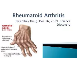
Introduction Rheumatoid arthritis (RA) is a chronic multisystemic disease of unknown cause. Characteristic feature: inflammatory synovitis - peripheral joints / symmetrical distribution - cartilage destruction / bone erosion - joint deformity After the metacarpophalangeal joints, the most common region to be involved in RAis the cervical spine. History: 1890, *Garrod reported that 36% of his pts with RA had c-spine involvement. * Garrod, A.Griffins Medical Series.1890. Available at: http://books.google. Accessed July 11, 2011.
Introduction Radiographic signs: 43-86% *Pellicci et al ( 5 yrs study / 106 RA pts) radiological evidence of c-spine involvement: 43% of pts / baseline 86% of pts / follow-up **Mikulowski et al: fatal cord compresion 10% in pts with RA Wasserman B,et al.Bull NYU Hospfor Jt Dis. 2011;69(2):136-48. * Pellicci P et al.J Bone Joint Surg Am. 1981;63:342-50. ** Mikulowski P et al. ActaMed Scand. 1975;198(6):445-51.
Introduction • Risk factors for c-spine involvement: – Males – RF - Rheumatoid nodules • – Peripheral activity • - Vasculitis • – Corticosteroid use • Clinical signs: • – Neck pain 40 to 88% • – Neurologic compromise 7 to 34% * Wasserman B,et al.Bull NYU Hospfor Jt Dis. 2011;69(2):136-48
RA & C-Spine Imaging • • Atlanto-axial subluxation( 65% of all cervicalsubluxations ) • - majority anterior • - 20% lateral • - 7%posterior • - rotatory rare • • Superior migration of the odontoid • - second most common deformity • - 20% of pts • - odontoid erosions • Subaxial c-spine involvement • - Subaxial subluxation :15% of pts • - Apophyseal joint ankylosis * Wasserman B,et al.Bull NYU Hospfor Jt Dis. 2011;69(2):136-48
RA & C-Spine Imaging Radiography • Anterior atlantoaxial subluxation • Vertical subluxation • Subaxial spinal involvement - Subaxial subluxation Magnetic Resonance Imaging • Pannus • Spinal cord
RA & C-Spine Imaging Radiography • Anterior atlantoaxial subluxation (AAS) AAS : 50% of pts symptomatic The role of plain radiography is to establish whether there are risk factors for cord compression.
AAS - Anterior atlantodental interval (AADI) AADI > 3-6 mm: early instability transverse lig. AADI > 6 mm transverse & alar lig. AADI > 9 mm surgical stabilization. AADI : yellow line
AAS Neutral Flexion AADI : yellow line
AAS - Posterior atlantodental interval (PADI) • All cervical spinal levels • cord: 10 mm • CSF: 2 mm • dura: 2 mm • PADI > 14 mm • (avoid cord compression) • spinal canal: 17-29 mm at C1 PADI : red line PADI : red line
AAS Neutral Flexion PADI : red line
Vertical subluxation McGregor´s line-Odontoid tip > 4.5 mm
Vertical subluxation Ranawat method (♂ > 15 mm & ♀ > 13 mm)
Vertical subluxation Clark’s stations
Vertical subluxation cervicomedullary angle (normalrange: 135° to 175°)
Subaxial subluxation Subluxation > 1mm > 3,5 mm !!! • Cervical Height Index (CHI) • - subluxations at multiple levels • - loss of disk height • bony collapse • CHI < 2(neurologic compromise)
Subaxial subluxation Zikou AK, et al.J Rheumatol 32: 801-806, 2005.
Sudaxial spinal involvement • Apophyseal joints ( erosions - ankylosis) • Intervertebral disk - space narrowing • Irregularity of the subchondral margins of the vertebral bodies • Erosion and sclerosis • Corticosteroid - ischemic necrosis of bone - vertebral collapse
RA & C-Spine Imaging Magnetic Resonance Imaging • Major indications for c-spine MRI in RA: • abnormal measurements on plain radiographs • unremitting suboccipital /cervical pain • progressive / severe subluxations • symptoms of cord/brainstem/vert. art. compression MRI : evaluation of the spinal cord and neural elements - Presence and effect of pannus on the spinal cord - Spinal cord signal can be assessed (edematous spinal cord changes: poor clinical status, poor prognosis & poor postoperative outcome)
AAS “pannus”
Odontoid erosions - “pannus” Zikou AK, et al. Clin Exp Rheumatol 23: 665-670, 2005.
Subaxial subluxation Zikou AK, et al. Clin Exp Rheumatol 23: 665-670, 2005.
AAS Subaxial subluxations
Take home messages Plain radiography : Flexion / extension views - the level of involvement - evidence of instability Further imaging with MRI : pannus & spinal cord AADI > 9 mm or PADI < 14 mm Vertical subluxation Subaxial subluxation > 3,5 mm The major role for MRI : pre & after operative assessment
* 165 RA pts ( 143♀/ 22♂) mean age: 59,6 ± 12,5 yrs disease duration: 12,3 ± 7,9 yrs RF (+) : 63,6% Radiological findings: 146 pts - AAS: 20,6% - Odontoid erosions: 2,4% - Sudaxial subluxations: 43,6% - Disk space narrowing: 66,1% - Vertebral plate erosions - sclerosis: 43,6% C - spine involvement: frequent finding mild severity ** 51 RA pts ( 42♀/ 9♂) mean age: 56,5 ± 10,4 yrs disease duration: 12,4 ± 8,5 yrs RF (+) : 64,7% clinical signs : c-pain & stiffness 30% Rx / MR findings: 40 / 44 pts - Peridental pannus: 88% - Odontoid erosions: 23,5% - AAS: 13,7% - Brainstem compression: 5,9% - Sudaxial subluxations: 10% Peridental pannus correlated (p<0,05) with: - DAS-28 - RF(+) - Erosive changes hand-wrist (Larsen criteria) *Zikou AK, et al.Radiological cervical spine involvement in patients with rheumatoid arthritis: a cross sectional study. J Rheumatol 32: 801-806, 2005. **Zikou AK, et al.Magnetic resonance imaging findings of the cervical spine in patients with rheumatoid arthritis: a cross sectional study.Clin Exp Rheumatol 23: 665-670, 2005.