Chapter 7: The Skeleton
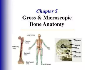
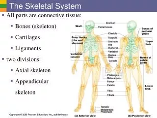
Chapter 7: The Skeleton. Part A. The Axial Skeleton. Consists of 80 bones Three major regions Skull Vertebral column Thoracic cage. The Axial Skeleton. Cranium. Skull. Facial bones. Clavicle. Thoracic cage (ribs and sternum). Scapula. Sternum. Rib. Humerus. Vertebra. Vertebral

Chapter 7: The Skeleton
E N D
Presentation Transcript
Chapter 7: The Skeleton Part A
The Axial Skeleton • Consists of 80 bones • Three major regions • Skull • Vertebral column • Thoracic cage
The Axial Skeleton Cranium Skull Facial bones Clavicle Thoracic cage (ribs and sternum) Scapula Sternum Rib Humerus Vertebra Vertebral column Radius Ulna Sacrum Carpals Phalanges Metacarpals Femur Patella Tibia Fibula Tarsals Metatarsals (a) Anterior view Phalanges Figure 7.1a
The Skull • Two sets of bones • Cranial bones • Enclose the brain in the cranial cavity • Gives attachment sites for head and neck muscles • Facial bones • Framework of face • Contains cavities for the special sense organs of sight, taste, and smell • Provides openings for the passage of air and food • Secures the teeth • Anchors the facial muscles of expression, which we use to show emotion
Bones of cranium (cranial vault) Coronal suture Squamous suture Facial bones Lambdoid suture (a) Cranial and facial divisions of the skull Figure 7.2a
Cranial Bones • Occipital bone • Parietal bones (2) • Frontal bone • Temporal bones (2) • Ethmoid bone • Sphenoid bone • Remember: • Old P-People From T-Texas Eat Spiders
Frontal bone Parietal bone Nasal bone Sphenoid bone (greater wing) Temporal bone Ethmoid bone Lacrimal bone Ethmoid bone Zygomatic bone Maxilla Mandible Vomer (a) Anterior view Mandibular symphysis Figure 7.4a
Parietal Bones and Major Associated Sutures • Superior and lateral aspects of cranial vault • Four sutures mark the articulations of parietal bones with frontal, occipital, and temporal bones: • Coronal suture—between parietal bones and frontal bone • Sagittal suture—between right and left parietal bones • Lambdoid suture—between parietal bones and occipital bone • Squamous (squamosal) sutures—between parietal and temporal bones on each side of skull
Frontal bone Coronal suture Sphenoid bone (greater wing) Parietal bone Ethmoid bone Temporal bone Lacrimal bone Lambdoid suture Squamous suture Nasal bone Occipital bone Zygomatic bone Zygomatic process Maxilla Occipitomastoid suture Mandible (a) External anatomy of the right side of the skull Figure 7.5a
Occipital Bone • Most of skull’s posterior wall and posterior cranial fossa • Contains the foramen magnum “large hole” through which the brain connects with the spinal cord • Articulates at the occipital condyles with 1st vertebra • Sites of attachment for the many neck and back muscles
Sagittal suture Parietal bone Sutural bone Lambdoid suture Occipital bone Occipitomastoid suture (b) Posterior view Figure 7.4b
Maxilla Intermaxillary suture Palatine bone Median palatine suture Maxilla Zygomatic bone Sphenoid bone (greater wing) Temporal bone (zygomatic process) Vomer Temporal bone Parietal bone Foramen magnum (a) Inferior view of the skull (mandible removed) Figure 7.6a
Temporal Bones • Inferolateral aspects of skull and parts of cranial floor • Contains the zygomatic process, external acoustic meatus, the styloid process, and the mastoid process • Articulates with the mandible at the TMJ
Sphenoid Bone • Complex, butterfly-shaped bone • Keystone bone • Articulates with all other cranial bones • Three pairs of processes • Contains the sella turcica and the hypophyseal fossa that surround the pituitary gland
Ethmoid Bone • Deepest skull bone • Superior part of nasal septum, roof of nasal cavities • Contributes to medial wall of orbits • Contains the superior and middle nasal conchae • Contains the crista galli (rooster’s comb) • The attachment site for the outermost covering of the brain
Sutural Bones • Tiny irregularly shaped bones that appear within sutures • http://www.sciencekids.co.nz/videos/humanbody/skullbones.html
Sagittal suture Parietal bone Sutural bone Lambdoid suture Occipital bone Occipitomastoid suture (b) Posterior view Figure 7.4b
Facial Bones (14 Total) Unpaired Bones: • Mandible • Vomer Paired Bones: • Maxillary bones (2) • Zygomatic bones (2) • Nasal bones (2) • Lacrimal bones (2) • Palatine bones (2) • Inferior nasal Conchae (2) Virgil Can Not Make My Pet Zebra Laugh!
Mandible • Lower jaw • Largest, strongest bone of face • Articulates at the temporomandibular joint (TMJ): only freely movable joint in skull
Temporomandibular joint Ramus of mandible Mandibular angle Body of mandible (a) Mandible, right lateral view Figure 7.11a
Maxillary Bones • Medially fused to form upper jaw and central portion of facial skeleton • Keystone bone of the facial bones: all facial bones except the mandible articulate with it
Zygomatic Bones • Cheekbones • Inferolateral margins of orbits • Articulates with 3 separate zygomatic processes • Frontal zygomatic process • Maxillary zygomatic process • Temporal zygomatic process
Nasal Bones and Lacrimal Bones • Nasal bones • Form bridge of nose • Attach to the cartilage that forms most of the skeleton of the nose • Lacrimal bones • In medial walls of orbits • Forms part of the canal that drains tears into the nasal cavity Lacrimation = crying/tear production
Frontal bone Parietal bone Nasal bone Sphenoid bone (greater wing) Temporal bone Ethmoid bone Lacrimal bone Ethmoid bone Zygomatic bone Maxilla Mandible Vomer (a) Anterior view Mandibular symphysis Figure 7.4a
Palatine Bones and Vomer • Palatine bones • Posterior one-third of hard palate • Posterolateral walls of the nasal cavity • Small part of the orbits • Vomer • Plow shaped • Lower part of nasal septum
Maxilla Intermaxillary suture Palatine bone Median palatine suture Maxilla Zygomatic bone Sphenoid bone (greater wing) Temporal bone (zygomatic process) Vomer Temporal bone Parietal bone Foramen magnum (a) Inferior view of the skull (mandible removed) Figure 7.6a
Orbits • Encase eyes and lacrimal glands • Sites of attachment for eye muscles • Formed by parts of seven bones • Frontal bones • Zygomatic • Sphenoid bones • Palatine • Ethmoid • Lacrimal • Maxilla Friendly Zebras Speed Past Elderly Lions Mating
Roof of orbit • Lesser wing ofsphenoid bone • Orbital plate offrontal bone Medial wall • Sphenoid body Lateral wall of orbit • Orbital plateof ethmoid bone • Zygomatic processof frontal bone • Greater wing ofsphenoid bone • Lacrimal bone • Orbital surface ofzygomatic bone Nasal bone Floor of orbit Zygomatic bone • Orbital surface ofmaxillary bone • Zygomatic bone (b) Contribution of each of the seven bones forming the right orbit Figure 7.13a
Nasal Cavity • Roof, lateral walls, and floor formed by parts of four bones • Ethmoid • Palatine bones • Maxillary bones • Inferior nasal conchae • Nasal septum of bone and hyaline cartilage • Ethmoid • Vomer • Anterior septal cartilage
Frontal sinus Ethmoid bone Nasal bone Maxillary bone (palatine process) Sphenoid bone Palatine bone (horizontal plate) Palatine bone (perpendicular plate) (a) Bones forming the left lateral wall of the nasal cavity (nasal septum removed) Figure 7.14a
Paranasal Sinuses • Mucosa-lined, air-filled spaces • Lighten the skull • Enhance resonance of voice • Found in frontal, sphenoid, ethmoid, and maxillary bones
Frontal sinus Frontal sinus Ethmoidal air cells (sinus) Ethmoidal air cells Sphenoid sinus Sphenoid sinus Maxillary sinus Maxillary sinus (b) Medial aspect (a) Anterior aspect Figure 7.15
Hyoid Bone • Not a bone of the skull • Does not articulate directly with another bone • Site of attachment for muscles of swallowing and speech
Developmental Aspects of the Skull • At birth, the newborn’s skull not fully developed and sutures • have not yet fused • Allows for head compression during birth • Allows for brain growth in the infant • Unossified regions are covered with fibrous membranes called • fontanelles “little fountains” • Anterior fontanelle is present until 1-1/2 –2 years of age
Homeostatic Imbalance of the Skull The Cleft Lip and Palate Caused by right and left halves of the palate failing to fuse medially Leads to difficulties feeding/nursing Risk for aspiration (inhalation) pneumonia