Download
1 / 21
210 likes | 656 Vues
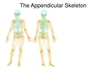
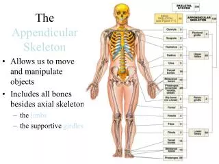
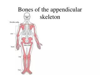
Bones of the appendicular skeleton. The Skeleton: Appendicular Skeleton. Acromio- clavicular joint. Clavicle. Scapula. Articulated pectoral girdle. Sternal (medial) end. Posterior. Anterior. Acromial (lateral) end. (b). Right clavicle, superior view. Figure 7.24b.

E N D
Acromio- clavicular joint Clavicle Scapula Articulated pectoral girdle
Sternal (medial) end Posterior Anterior Acromial (lateral) end (b) Right clavicle, superior view Figure 7.24b
Suprascapular notch Acromion Superior border Coracoid process Superior angle Glenoid cavity Subscapular fossa Lateral border Medial border Inferior angle (a) Right scapula, anterior aspect Figure 7.25a
Coracoid process Suprascapular notch Superior angle Acromion Supraspinous fossa Glenoid cavity at lateral angle Spine Infraspinous fossa Medial border Lateral border (b) Right scapula, posterior aspect
Supraspinous fossa Supraglenoid tubercle Acromion Coracoid process Glenoid cavity Spine Supraspinous fossa Infraglenoid tubercle Infraspinous fossa Infraspinous fossa Subscapular fossa Subscapular fossa Posterior Anterior (c) Right scapula, lateral aspect Inferior angle
Humerus Greater tubercle Head of humerus Lesser tubercle Anatomical neck Inter- tubercular sulcus Deltoid tuberosity Lateral supracondylar ridge Coronoid fossa Radial fossa Medial epicondyle Capitulum Trochlea (a) Anterior view
Radial notch of the ulna Olecranon process Trochlear notch Head Head of radius Coronoid process Neck Radial tuberosity Neck of radius Proximal radioulnar joint Interosseous membrane Ulna Radius Ulnar notch of the radius Radius Head of ulna Styloid process of ulna Styloid process of radius Distal radioulnar joint Styloid process of radius (a) Anterior view (b) Posterior view
Olecranon process View Trochlear notch Coronoid process Radial notch (c) Proximal portion of ulna, lateral view Ulnar notch of radius Articulation for lunate Articulation for scaphoid Styloid process Styloid process Head of ulna View (d) Distal ends of the radius and ulna at the wrist
Coronoid fossa Humerus Capitulum Medial epicondyle Trochlea Head of radius Coronoid process of ulna Radial tuberosity Radial notch Radius Ulna (c) Anterior view at the elbow region Olecranon fossa Humerus Olecranon process Lateral epicondyle Medial epicondyle Head Ulna Neck Radius (d) Posterior view of extended elbow
Phalanges • Distal • Middle • Proximal Metacarpals • Head • Shaft • Base Sesamoid bones Carpals Carpals Carpals • Trapezium • Hamate • Trapezium • Trapezoid • Capitate • Trapezoid • Scaphoid • Pisiform • Scaphoid • Triquetrum Radius • Lunate Ulna Radius (a) Anterior view of left hand (b) Posterior view of left hand Recommend a mnemonic device here: SLTPTTCH
Pelvic (Hip) Girdle • Two hip bones (each also called coxal bone or os coxae) • Attach the lower limbs to the axial skeleton with strong ligaments • Transmit weight of upper body to lower limbs • Support pelvic organs • Each hip bone consists of three fused bones: ilium, ischium, and pubis • Together with the sacrum and the coccyx, these bones form the bony pelvis
Base of sacrum Iliac crest Sacroiliac joint Iliac fossa Anterior superior iliac spine Sacral promontory Coxal bone (os coxae or hip bone) Anterior inferior iliac spine llium Sacrum Pubic bone Pelvic brim Coccyx Acetabulum Pubic tubercle Ischium Pubic crest Pubic symphysis Pubic arch
Anterior gluteal line Ilium Posterior gluteal line Iliac crest Posterior superior iIiac spine Anterior superior iliac spine Posterior inferior iliac spine Inferior gluteal line Greater sciatic notch Anterior inferior iliac spine Ischial body Acetabulum Ischial spine Pubic body Lesser sciatic notch Pubis Ischium Inferior ramus of pubis Obturator foramen Ischial ramus (a) Lateral view, right hip bone
Ilium Iliac fossa Iliac crest Posterior superior iliac spine Anterior superior iliac spine Posterior inferior iliac spine Anterior inferior iliac spine Auricular surface Body of the ilium Arcuate line Greater sciatic notch Superior ramus of pubis Ischial spine Lesser sciatic notch Pubic tubercle Obturator foramen Articular surface of pubis (at pubic symphysis) Ischium Ischial ramus Inferior ramus of pubis (b) Medial view, right hip bone Figure 7.30b
Neck Fovea capitis Greater trochanter Head Inter- trochanteric crest Lesser trochanter Intertrochanteric line Gluteal tuberosity Linea aspera Apex Anterior Facet for lateral condyle of femur Facet for medial condyle of femur Lateral condyle Medial and lateral supra- condylar lines Lateral epicondyle Surface for patellar ligament Intercondylar fossa Medial condyle Posterior Adductor tubercle (a) Patella (kneecap) Lateral epicondyle Medial epicondyle Patellar surface Anterior view Posterior view (b) Femur (thigh bone)
Lateral condyle Intercondylar eminence Head Medial condyle Proximal tibiofibular joint Tibial tuberosity Interosseous membrane Anterior border Fibula Tibia Distal tibiofibular joint Articular surface Lateral malleolus Medial malleolus (a) Anterior view
Articular surface of medial condyle Articular surface of lateral condyle Medial condyle Head of fibula Interosseous membrane Tibia Fibula Articular surface Medial malleolus Lateral malleolus (b) Posterior view Figure 7.32b
Phalanges Distal Middle Proximal 1 2 3 4 5 Metatarsals Medial cuneiform Intermediate cuneiform Lateral cuneiform Navicular Cuboid Tarsals Talus Trochlea of talus Calcaneus (a) Superior view The new milccurdled