The Appendicular Skeleton
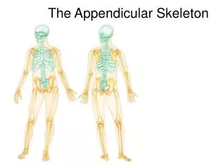
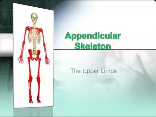
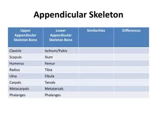
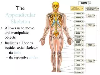
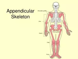
The Appendicular Skeleton. The Appendicular Skeleton. These consist of the limbs and their girdles. Their major function is to carry out movement. The Appendicular Skeleton. These consist of the limbs and their girdles. Their major function is to carry out movement.


The Appendicular Skeleton
E N D
Presentation Transcript
The Appendicular Skeleton These consist of the limbs and their girdles. Their major function is to carry out movement.
The Appendicular Skeleton These consist of the limbs and their girdles. Their major function is to carry out movement.
The Appendicular Skeleton The pectoral girdle consists of the clavicle and scapula. 1) Only the clavicle attaches to the axial skeleton (sternal end) 2) The scapula is designed for flexibility
Figure 7.24a The pectoral girdle and clavicle. Acromio- clavicular joint Clavicle Scapula (a) Articulated pectoral girdle
Figure 7.24b The pectoral girdle and clavicle. Sternal (medial) end Posterior Anterior Acromial (lateral) end (b) Right clavicle, superior view
The Appendicular Skeleton The scapula has three borders. • Superior border • Medial border which lies toward the vertebral column • The lateralborder which has the glenoid cavity
Figure 7.25a The scapula. Suprascapular notch Acromion Superior border Coracoid process Superior angle Glenoid cavity Subscapular fossa Lateral border Medial border Inferior angle (a) Right scapula, anterior aspect
Figure 7.25b The scapula. Coracoid process Suprascapular notch Superior angle Acromion Supraspinous fossa Glenoid cavity at lateral angle Spine Infraspinous fossa Medial border Lateral border (b) Right scapula, posterior aspect
Figure 7.25c The scapula. Supraspinous fossa Supraglenoid tubercle Acromion Coracoid process Glenoid cavity Spine Supraspinous fossa Infraglenoid tubercle Infraspinous fossa Infraspinous fossa Subscapular fossa Subscapular fossa Posterior Anterior (c) Right scapula, lateral aspect Inferior angle
The Appendicular Skeleton The humerus articulates with the scapula at the shoulder and the ulna and radius distally. The proximal head has the greater and lesser tubercles and anatomical neck which is where the rotator cuff muscles attach.
Figure 7.26a The humerus of the right arm and detailed views of articulation at the elbow. Greater tubercle Head of humerus Lesser tubercle Anatomical neck Inter- tubercular sulcus Deltoid tuberosity Lateral supracondylar ridge Coronoid fossa Radial fossa Medial epicondyle Capitulum Trochlea (a) Anterior view
The Appendicular Skeleton Just distally is the surgical neck, the most frequently fractured portion of the humerus.
The Appendicular Skeleton • The deltoid tuberosity on the lateral side is for the attachment of the deltoid muscle • Distally there are two condyles, the medical trochlea which articulates with the ulna and the lateral capitulum which articulates with the radius.
Figure 7.26c The humerus of the right arm and detailed views of articulation at the elbow. Humerus Coronoid fossa Medial epicondyle Capitulum Trochlea Head of radius Coronoid process of ulna Radial tuberosity Radial notch Radius Ulna (c) Anterior view at the elbow region
Telling Left from Right • Orient the bone so that the rounded head is superior (up) and pointing medially. • Look for the deep olecranon fossa on the posterior side.
The Appendicular Skeleton What is the medial epicondyle famous for?
The Appendicular Skeleton What is the medial epicondyle famous for? The Funny Bone
The Funny Bone The ulnar nerve is the largest unprotected nerve in the human body unprotected by muscle or bone), so injury is common.
The Funny Bone This nerve is directly connected to the little finger, and the adjacent half of the ring finger, supplying the palmar side of these fingers, including both front and back of the tips.
The Funny Bone The clawed hand can be a result of ulnar nerve damage.
The Appendicular Skeleton The ulna and radial bones form the distal lower limb. The ulna is medial and the radius is lateral.
The Appendicular Skeleton The olecranon process (elbow) and the coronoid processes are the major land marks on the proximal portion of the ulna. The ulna plays no major role in wrist movement. Its only action is extension and flexion of the lower limb.
Figure 7.27c Radius and ulna of the right forearm. Olecranon process View Trochlear notch Coronoid process Radial notch (c) Proximal portion of ulna, lateral view
The Appendicular Skeleton The radius is shaped like the head of a nail. Its head is concave. Its distal end is highlighted by the styloid process. The radius allows for pronation and supination of the wrist.
Telling Left from Right • Place the ulna so that the trochlear notch faces up, the radial notch should face medially. • Place the radius so the distal styloid process is lateral and the radial tuberosity is medial
The Hand The “hand” is composed of 8 carpals and 5 metacarpals. Distally are the phalanges, these begin at the knuckles.
Figure 7.28 Bones of the left hand. Phalanges • Distal • Middle • Proximal Metacarpals • Head • Shaft • Base Sesamoid bones Carpals Carpals Carpals • Trapezium • Hamate • Trapezium • Trapezoid • Capitate • Trapezoid • Scaphoid • Pisiform • Scaphoid • Triquetrum Radius • Lunate Ulna Radius (a) Anterior view of left hand (b) Posterior view of left hand
The Hand Carpal tunnel syndrome is pain, tingling, and other problems in your hand because of pressure on the median in your wrist. It is a common repetitive injury.
The Appendicular SkeletonThe Pelvic Girdle The pelvic girdle attaches the lower limbs to the axial skeleton. The hip is also known as the os coxae It is made up of three separate bones: • Ischium • Ilium & • Pubis
Figure 7.29 Articulated pelvis showing the two hip (coxal) bones (which together form the pelvic girdle), the sacrum, and the coccyx. Base of sacrum Iliac crest Sacroiliac joint Iliac fossa Anterior superior iliac spine Sacral promontory Coxal bone (os coxae or hip bone) Anterior inferior iliac spine llium Sacrum Pubic bone Pelvic brim Coccyx Acetabulum Pubic tubercle Ischium Pubic crest Pubic symphysis Pubic arch
Figure 7.30c Bones of the bony pelvis. Anterior gluteal line Ilium Posterior gluteal line Anterior superior iliac spine Posterior superior iliac spine Anterior inferior iliac spine Inferior gluteal line Posterior inferior iliac spine Acetabulum Greater sciatic notch Ischial body Pubic body Ischial spine Pubic tubercle Lesser sciatic notch Ischium Inferior ramus of pubis Ischial tuberosity Ischial ramus Obturator foramen (c) Lateral view, right hip bone
Dimples of Venus These are indentations sometimes visible on the human lower back, just superior to the gluteal cleft. They are directly superficial to the two sacroiliac joints, the sites where the sacrum attaches to the ilium of the pelvis.
The Patella The patella is a triangular, sesamoid bone enclosed in the quadriceps tendon. It helps to improve leverage of the thigh muscles on the tibia.
Figure 7.31a Bones of the right knee and thigh. Apex Anterior Facet for lateral condyle of femur Facet for medial condyle of femur Surface for patellar ligament Posterior (a) Patella (kneecap)
Dislocation of the Patella • Kneecap (patella) dislocation is often seen in women. • It usually occurs after a sudden change in direction when your leg is planted. This puts your kneecap under stress.
Dislocation of the Patella • Dislocation may also occur as a direct result of injury. When the kneecap is dislocated, it can slip sideways and around to the outside of the knee.
The Femur The femur is the longest and strongest bone of the body.
The Femur The femur is the longest and strongest bone of the body. Its identified by having a large and have a distinct rounded head
The Femur The femur has a distinct neck separating the head from the rest of the bone.